

The applicability of furfuryl-gelatin as a novel bioink for tissue engineering applications
- PMID: 29656592
- PMCID: PMC6188846
- DOI: 10.1002/jbm.b.34123
The applicability of furfuryl-gelatin as a novel bioink for tissue engineering applications
Abstract
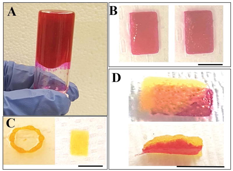
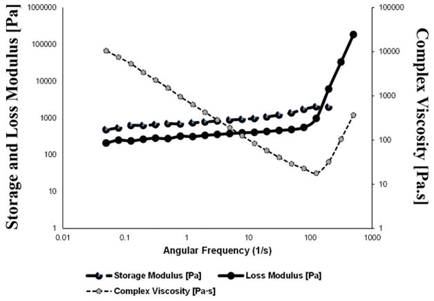
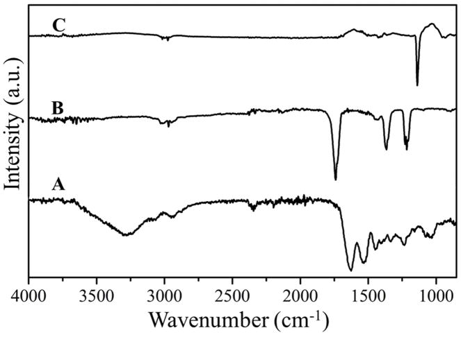
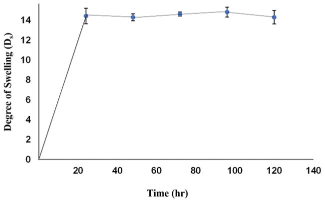
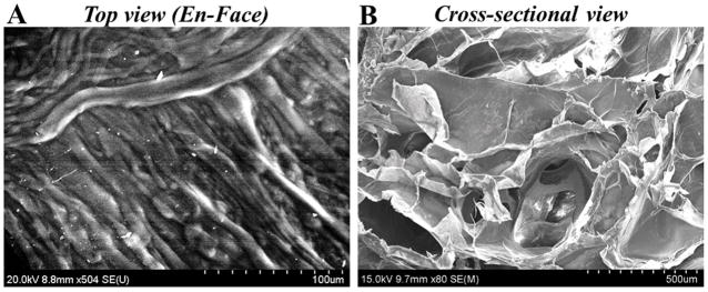
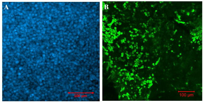
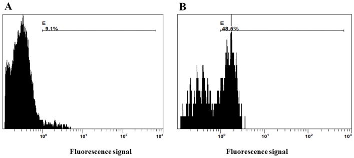
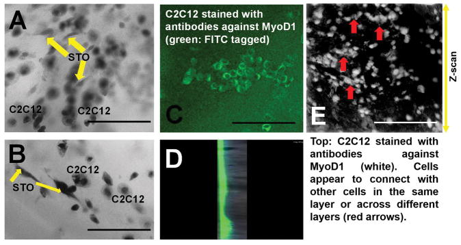
Three-dimensional bioprinting is an innovative technique in tissue engineering, to create layer-by-layer structures, required for mimicking body tissues. However, synthetic bioinks do not generally possess high printability and biocompatibility at the same time. So, there is an urgent need for naturally derived bioinks that can exhibit such optimized properties. We used furfuryl-gelatin as a novel, visible-light crosslinkable bioink for fabricating cell-laden structures with high viability. Hyaluronic acid was added as a viscosity enhancer and either Rose Bengal or Riboflavin was used as a visible-light crosslinker. Crosslinking was done by exposing the printed structure for 2.5 min to visible light and confirmed using Fourier transform infrared spectroscopy and rheometry. Scanning electron microscopy revealed a highly porous networked structure. Three different cell types were successfully bioprinted within these constructs. Mouse mesenchymal stem cells printed within monolayer and bilayer sheets showed viability, network formation and proliferation (∼5.33 times) within 72 h of culture. C2C12 and STO cells were used to print a double layered structure, which showed evidence of the viability of both cells and heterocellular clusters within the construct. This furfuryl-gelatin based bioink can be used for tissue engineering of complex tissues and help in understanding how cellular crosstalk happens in vivo during normal or diseased pathology. © 2018 Wiley Periodicals, Inc. J Biomed Mater Res Part B: Appl Biomater, 107B: 314-323, 2019.
Keywords: bilayer sheets; biocompatibility; furfuryl-gelatin; hyaluronic acid; visible-light crosslinkable bioink.
© 2018 Wiley Periodicals, Inc.
Figures
References
-
- Murphy SV, Atala A. 3D bioprinting of tissues and organs. Nature biotechnology. 2014;32(8):773–785. - PubMed
-
- Yan Y, et al. Fabrication of viable tissue-engineered constructs with 3D cell-assembly technique. Biomaterials. 2005;26(29):5864–5871. - PubMed
-
- Ferris CJ, et al. Bio-ink for on-demand printing of living cells. Biomaterials Science. 2013;1(2):224–230. - PubMed
-
- Drury JL, Mooney DJ. Hydrogels for tissue engineering: scaffold design variables and applications. Biomaterials. 2003;24(24):4337–4351. - PubMed
-
- Kang H-W, Tabata Y, Ikada Y. Fabrication of porous gelatin scaffolds for tissue engineering. Biomaterials. 1999;20(14):1339–1344. - PubMed
Publication types
MeSH terms
Substances
Grants and funding
LinkOut - more resources
Full Text Sources
Other Literature Sources
