

doi: 10.1021/jacs.8b01236.
Epub 2018 May 7.
Direct Experimental Characterization of Glycosyl Cations by Infrared Ion Spectroscopy
Affiliations
- PMID: 29656643
- PMCID: PMC5958338
- DOI: 10.1021/jacs.8b01236
Item in Clipboard
Direct Experimental Characterization of Glycosyl Cations by Infrared Ion Spectroscopy
J Am Chem Soc.
.
Abstract
Glycosyl cations are crucial intermediates formed during enzymatic and chemical glycosylation. The intrinsic high reactivity and short lifetime of these reaction intermediates make them very challenging to characterize using spectroscopic techniques. Herein, we report the use of collision induced dissociation tandem mass spectrometry to generate glycosyl cations in the gas phase followed by infrared ion spectroscopy using the FELIX infrared free electron laser. The experimentally observed IR spectra were compared to DFT calculated spectra enabling the detailed structural elucidation of elusive glycosyl oxocarbenium and dioxolenium ions.
Conflict of interest statement
The authors declare no competing financial interest.
Figures
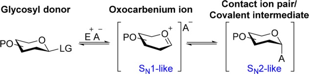
LG = Leaving group, E = Electrophile, A =
Anion, P = Protecting group.

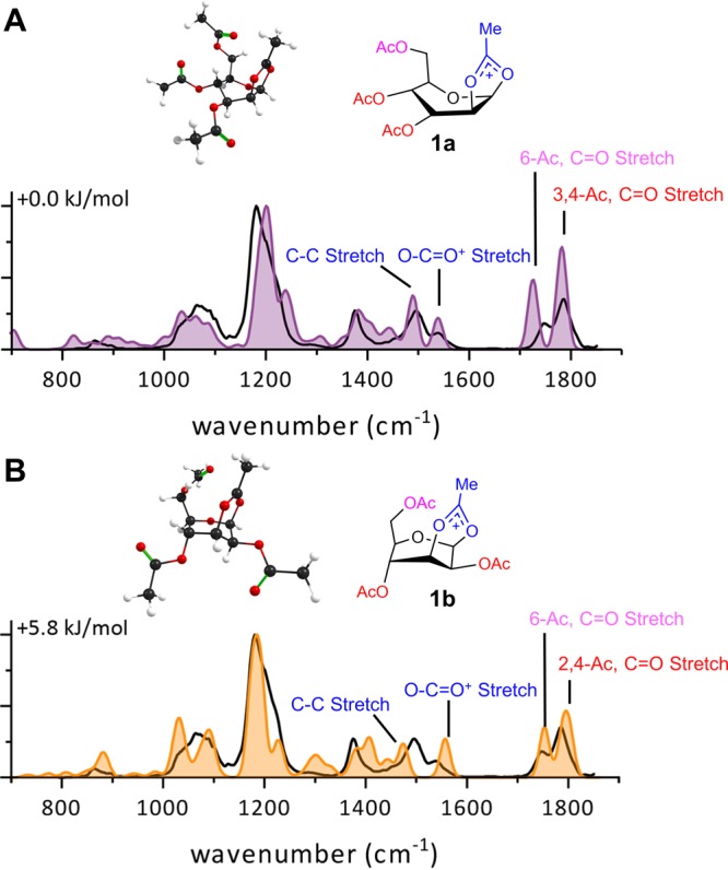
Comparison of the calculated spectra (filled)
of 2-O-acetyl- (1a) or 3-O-acetyl- (1b) participation with the measured IR ion
spectrum of mannosyl
cation derived from 1 (black line in both panels).

Comparison of the calculated spectra (filled) of the 3E (2a) or 4H3 (2b) oxocarbenium ion conformer
with the
measured IR ion spectrum of the m/z = 219 CID fragment of 2 (black line in both panels).
Energies are relative to the 0.0 kJ/mol structure as reported in Figure S6 .
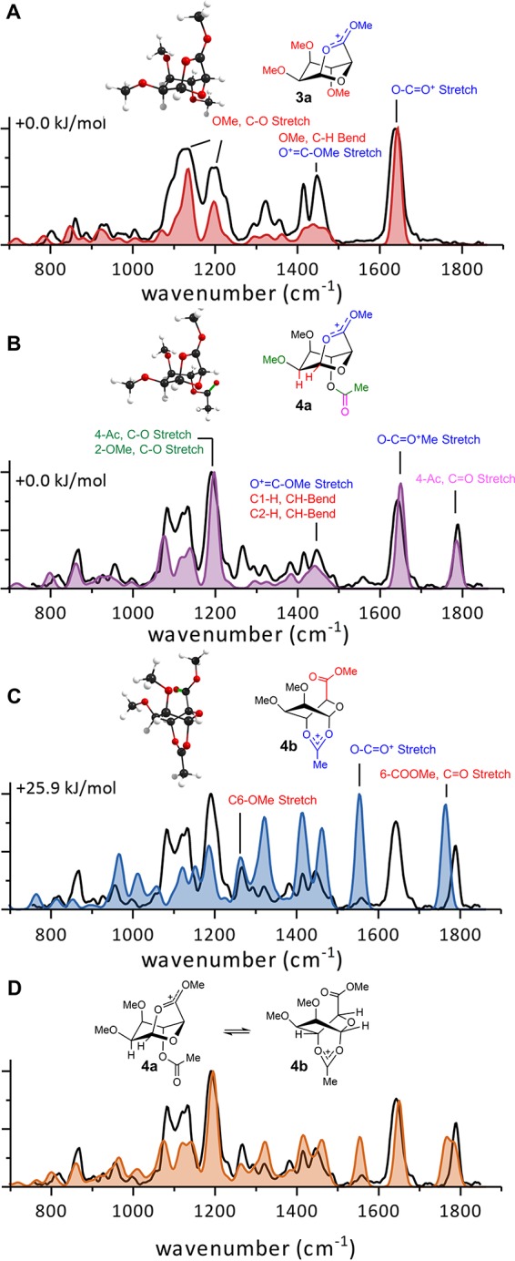
Comparison
of calculated spectra (filled) with the measured IR
ion spectra (black line) of mannuronic acids 3a, 4a, 4b (A–C) and a mixture of isomers 4a and 4b (D).
References
-
- Horenstein N. A. In Advances in Physical Organic Chemistry; Richard J. P., Ed.; Elsevier: New York, 2006; Vol. 41, p 275.
Publication types
LinkOut - more resources
Full Text Sources
Other Literature Sources
