. 2018 May 2;98(3):521-529.e3.
doi: 10.1016/j.neuron.2018.03.034. Epub 2018 Apr 12.

Structural Mechanism of Functional Modulation by Gene Splicing in NMDA Receptors

Affiliations

Structural Mechanism of Functional Modulation by Gene Splicing in NMDA Receptors

Michael C Regan et al. Neuron. .

Abstract

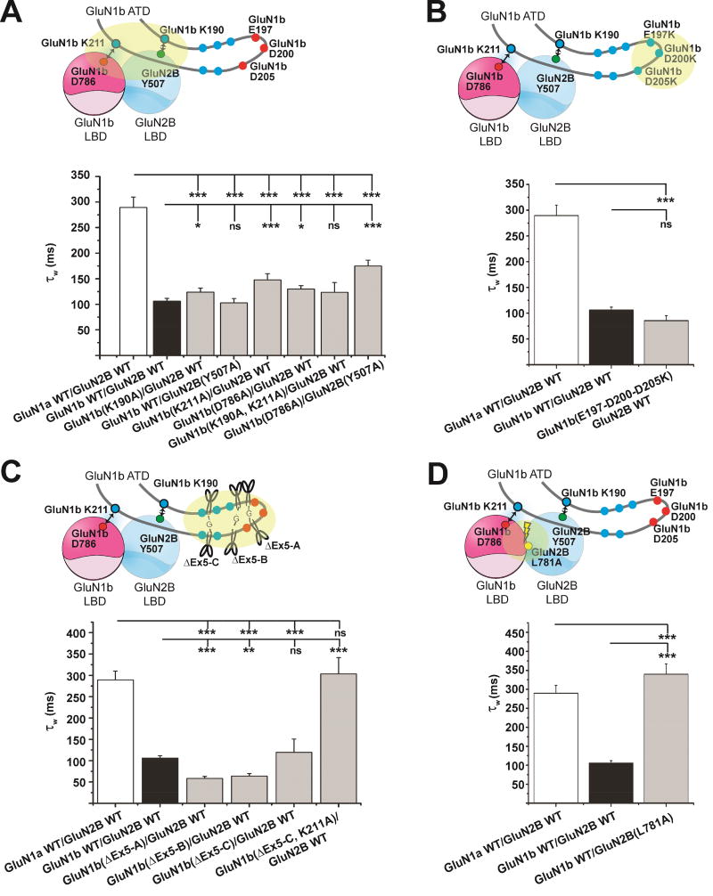
Alternative gene splicing gives rise to N-methyl-D-aspartate (NMDA) receptor ion channels with defined functional properties and unique contributions to calcium signaling in a given chemical environment in the mammalian brain. Splice variants possessing the exon-5-encoded motif at the amino-terminal domain (ATD) of the GluN1 subunit are known to display robustly altered deactivation rates and pH sensitivity, but the underlying mechanism for this functional modification is largely unknown. Here, we show through cryoelectron microscopy (cryo-EM) that the presence of the exon 5 motif in GluN1 alters the local architecture of heterotetrameric GluN1-GluN2 NMDA receptors and creates contacts with the ligand-binding domains (LBDs) of the GluN1 and GluN2 subunits, which are absent in NMDA receptors lacking the exon 5 motif. The unique interactions established by the exon 5 motif are essential to the stability of the ATD/LBD and LBD/LBD interfaces that are critically involved in controlling proton sensitivity and deactivation.

Keywords: NMDA receptor; alternative splicing; cryo-EM; electrophysiology.


Conflict of interest statement

Declaration of Interests

Authors declare no conflict of interests.

Figures

Figure 1
Figure 1. Inclusion of Exon 5 in GluN1 alters ion channel properties in NMDA receptors
Two-electrode voltage clamp recording from cRNA injected Xenopus oocytes (A) and fast-perfusion whole cell patch clamp recording from DNA transfected HEK293 cells (B) of the GluN1-4a-GluN2B (−Exon 5) and GluN1-4b-GluN2B (+Exon 5) NMDA receptors illustrating reduced proton sensitivity and faster deactivation speed elicited by Exon 5, respectively. Error bars in (A) represent ±SD for data obtained from 15 and 17 different oocytes for the −Exon 5 and +Exon 5, respectively. The recording traces in (B) are fit (magenta and red) by a double exponential equation.
Figure 2
Figure 2. Construct design and protein purification
(A) GluN1b and GluN2B constructs used in the current structural study (GluN1b-GluN2BEMX). The glycosylation knock-out mutations are to improve the expression level (green ovals). Free cysteines in GluN2B were mutated to serines (orange ovals). Cross-link mutations were incorporated to reduce conformational heterogeneity and to improve the quality of 3D reconstruction (gray ovals). (B) SDS-PAGE (8%) gel and the chromatograph from size-exclusion chromatography (Superose-6) showing purity and size-homogeneity of the GluN1b-GluN2BEMX sample.
Figure 3
Figure 3. Cryo-electron microscopy structure identifies the Exon 5 encoded motif at domain and subunit interfaces
(A) Cryo-EM structure of the intact GluN1b-GluN2B NMDA receptor at ~4.6 Å. The GluN1b subunits are colored magenta and gold while the GluN2B subunits are colored cyan and blue. (B) Highlighted region from (A), illustrating cryo-EM density for residues of the Exon 5 motif at the ATD/LBD interface. (C) Overlay of models from the GluN1a-GluN2B and GluN1b-GluN2B intact NMDA receptors. Note the large shift in position of GluN1 Lys190 between the GluN1a and GluN1b isoforms.
Figure 4
Figure 4. Assessment of interdomain and intersubunit interactions mediated by the Exon 5 motif
(A) Model of the GluN1b (magenta) and GluN2B (cyan) subunits with key residues labeled. We identified three groups of interactions at this site (I–III) through crosslinking and Western blot analysis. (B) Results of Western blots after crosslinking experiments for our series of mutants. Single cysteine mutants were unable to form dimers, while pairs of mutants were detected at masses indicating disulfide bond formation, which could be eliminated by application of a reducing agent.
Figure 5
Figure 5. Interactions between the Exon 5 motif and the LBDs control proton sensitivity
Here, we employed four types of mutations in the context of TEVC experiments to explore several potential effects of the Exon 5 motif: (A) disruption the direct Exon 5 – LBD interactions, (B) Reversal of negative charges to positives, (C) alter local structure of the Exon 5 motif by truncations (detailed in Table S1), and (D) disruption of the heterodimeric interface between the GluN1 and GluN2B LBDs. Shown in gray lines and two-tone spheres are the Exon 5 motif and the GluN1b-GluN2B LBD heterodimers, respectively. The lightning bolt and scissors indicate disruption of interactions and truncations, respectively. The areas of engineered mutations are highlighted by transparent yellow ovals. IC50 values of proton inhibition for the wild type GluN1-4a-GluN2B and GluN1-4b-GluN2B NMDA receptors and the mutant GluN1-4b-GluN2B NMDA receptors were estimated by recording macroscopic current at various pHs by TEVC from at least five oocytes per construct. (*: p < 0.05, **: p < 0.01; ***: p < 0.001; ns: not significant, as determined by one-way ANOVA with Tukey’s test. Bar graphs represent mean ± SEM). All of the TEVC recordings look similar to the ones in Figure 1A.
Figure 6
Figure 6. Structure and proper placement of the Exon 5 motif at the ATD-LBD interface is critical for controlling deactivation rates
The same set of the mutant GluN1-4b-GluN2B NMDA receptors in Figure 4 were tested for deactivation rates in HEK 293T cells by WCPC. No pronounced effects were observed by disrupting the Exon 5 – LBD interactions (A) or reversing residue charges (B). (C) To reverse the effect of Exon 5, a combination of truncation and disruption of the Exon 5 – LBD interaction (GluN1b ΔEx5-C/K211A) is necessary. (D) Disruption of the GluN1b-GluN2B LBD heterodimer interface by GluN2B Leu781Ala overrode the effect of Exon 5. In all cases, WCPC recordings on transfected HEK293 cells were done on at least six cells per construct. (*: p < 0.05, **: p < 0.01; ***: p < 0.001; ns: not significant, as determined by one-way ANOVA with Tukey’s test. Bar graphs represent mean ± SEM). Representative WCPC recordings are shown in Figure S5.

References

    1. Adams DR, Yuan H, Holyoak T, Arajs KH, Hakimi P, Markello TC, Wolfe LA, Vilboux T, Burton BK, Fajardo KF, et al. Three rare diseases in one Sib pair: RAI1, PCK1, GRIN2B mutations associated with Smith-Magenis Syndrome, cytosolic PEPCK deficiency and NMDA receptor glutamate insensitivity. Molecular genetics and metabolism. 2014;113:161–170. - PMC - PubMed
    1. Afonine PV, Headd JJ, Terwilliger TC, Adams PD. Computational Crystallography Newsletter. 2013;4:43–44.
    1. Bassler EL, Ngo-Anh TJ, Geisler HS, Ruppersberg JP, Grunder S. Molecular and functional characterization of acid-sensing ion channel (ASIC) 1b. J Biol Chem. 2001;276:33782–33787. - PubMed
    1. Castiglioni AJ, Raingo J, Lipscombe D. Alternative splicing in the C-terminus of CaV2.2 controls expression and gating of N-type calcium channels. J Physiol. 2006;576:119–134. - PMC - PubMed
    1. Chen C, Okayama H. High-efficiency transformation of mammalian cells by plasmid DNA. Molecular and cellular biology. 1987;7:2745–2752. - PMC - PubMed

Publication types

Substances