. 2018 Apr 19;173(3):595-610.e11.
doi: 10.1016/j.cell.2018.03.043. Epub 2018 Apr 12.

Deterministic Evolutionary Trajectories Influence Primary Tumor Growth: TRACERx Renal

Samra Turajlic  1 Hang Xu  2 Kevin Litchfield  2 Andrew Rowan  2 Stuart Horswell  3 Tim Chambers  2 Tim O'Brien  4 Jose I Lopez  5 Thomas B K Watkins  2 David Nicol  6 Mark Stares  2 Ben Challacombe  4 Steve Hazell  7 Ashish Chandra  8 Thomas J Mitchell  9 Lewis Au  10 Claudia Eichler-Jonsson  2 Faiz Jabbar  2 Aspasia Soultati  11 Simon Chowdhury  11 Sarah Rudman  11 Joanna Lynch  10 Archana Fernando  4 Gordon Stamp  12 Emma Nye  12 Aengus Stewart  3 Wei Xing  13 Jonathan C Smith  13 Mickael Escudero  3 Adam Huffman  13 Nik Matthews  14 Greg Elgar  14 Ben Phillimore  14 Marta Costa  14 Sharmin Begum  14 Sophia Ward  15 Max Salm  3 Stefan Boeing  3 Rosalie Fisher  2 Lavinia Spain  10 Carolina Navas  2 Eva Grönroos  2 Sebastijan Hobor  2 Sarkhara Sharma  2 Ismaeel Aurangzeb  2 Sharanpreet Lall  11 Alexander Polson  8 Mary Varia  8 Catherine Horsfield  8 Nicos Fotiadis  16 Lisa Pickering  10 Roland F Schwarz  17 Bruno Silva  13 Javier Herrero  18 Nick M Luscombe  19 Mariam Jamal-Hanjani  20 Rachel Rosenthal  21 Nicolai J Birkbak  22 Gareth A Wilson  22 Orsolya Pipek  23 Dezso Ribli  23 Marcin Krzystanek  24 Istvan Csabai  23 Zoltan Szallasi  25 Martin Gore  10 Nicholas McGranahan  20 Peter Van Loo  26 Peter Campbell  27 James Larkin  28 Charles Swanton  29 TRACERx Renal Consortium
Affiliations

Deterministic Evolutionary Trajectories Influence Primary Tumor Growth: TRACERx Renal

Samra Turajlic et al. Cell. .

Abstract

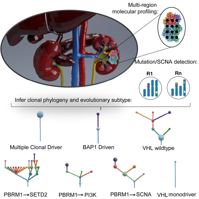
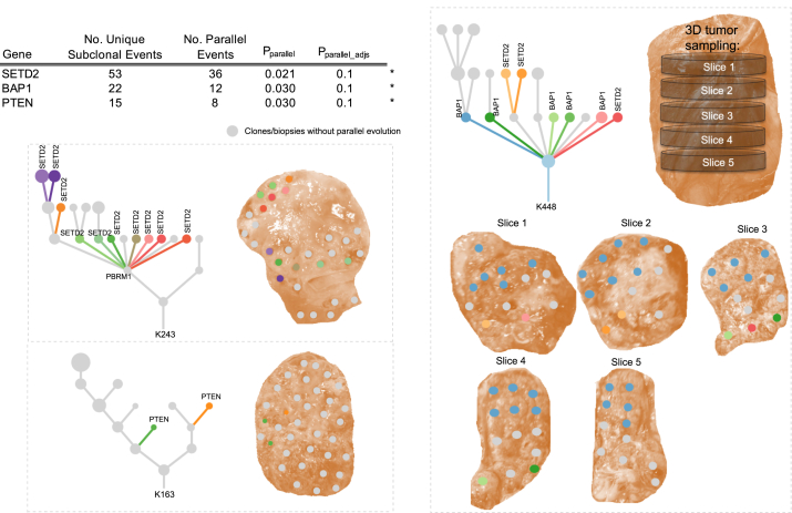
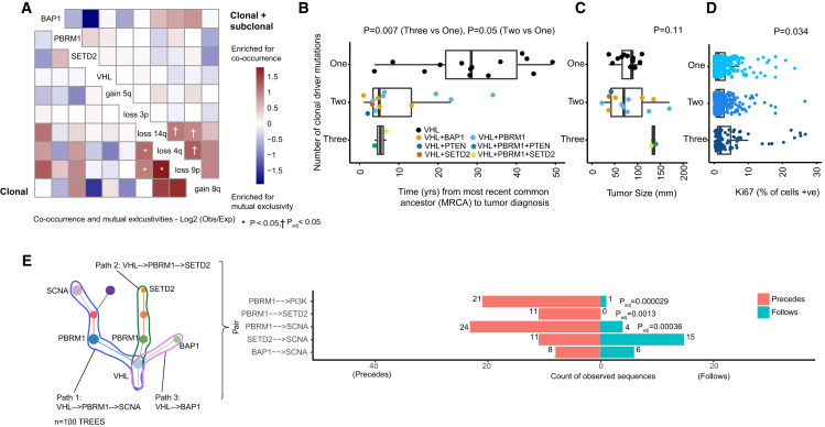
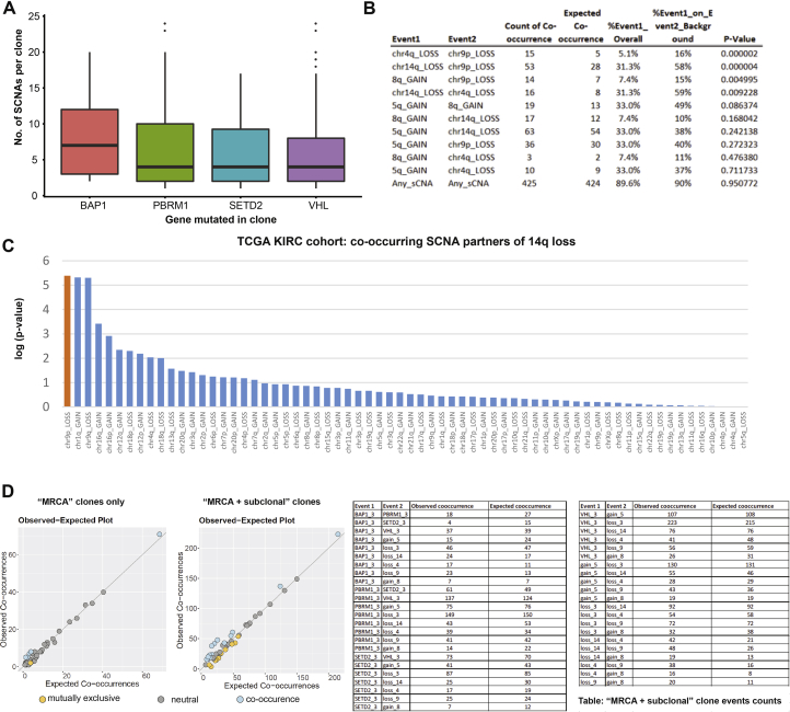
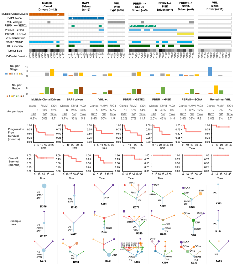
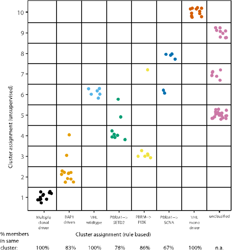
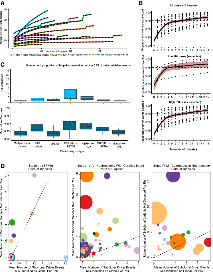
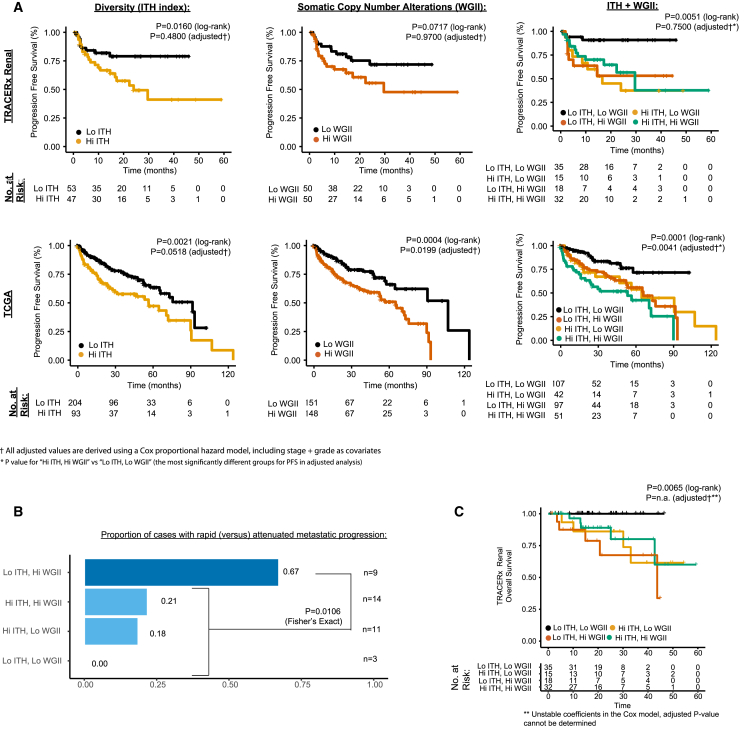
The evolutionary features of clear-cell renal cell carcinoma (ccRCC) have not been systematically studied to date. We analyzed 1,206 primary tumor regions from 101 patients recruited into the multi-center prospective study, TRACERx Renal. We observe up to 30 driver events per tumor and show that subclonal diversification is associated with known prognostic parameters. By resolving the patterns of driver event ordering, co-occurrence, and mutual exclusivity at clone level, we show the deterministic nature of clonal evolution. ccRCC can be grouped into seven evolutionary subtypes, ranging from tumors characterized by early fixation of multiple mutational and copy number drivers and rapid metastases to highly branched tumors with >10 subclonal drivers and extensive parallel evolution associated with attenuated progression. We identify genetic diversity and chromosomal complexity as determinants of patient outcome. Our insights reconcile the variable clinical behavior of ccRCC and suggest evolutionary potential as a biomarker for both intervention and surveillance.

Trial registration: ClinicalTrials.gov NCT03226886.

Keywords: branched evolution; cancer evolution; chromosome instability; deterministic evolution; intratumor heterogeneity; linear evolution; metastasis; punctuated evolution; renal cell cancer; tumor diversity.


Figures

None
Graphical abstract
Figure S1
Figure S1
Consort Diagram, Related to STAR Methods (A and B) (A) shows the Consort diagram for the filtering steps leading to the reported cohort; (B) shows the summary of Driver Panel, Whole Exome and Whole Genome Sequencing in the TRACERx Renal 101 Cohort.
Figure 1
Figure 1
Overview (A) Overview of somatic driver alterations, including SNVs, DNVs, INDELs, and SCNAs, detected in the tumors of 101 TRACERx Renal cases. Rectangles and triangles indicate clonal and subclonal alterations, respectively. Parallel evolution is indicated in orange with a split indicating 2 or more parallel events. Five bilateral/multi-focal cases are shown on the right; distinct VHL mutations within tumor pairs are indicated with an asterisk. (B) Mutational frequency in 14 key driver genes in the TRACERx Renal cohort and three single biopsy ccRCC studies (TCGA KIRC, Sato et al. [2013], and Scelo et al. [2014]). Clonal mutations are shown in the darker shade, subclonal in lighter. (C) Frequency of SCNAs in the TRACERx Renal cohort. Copy number gains and losses are indicated in red and blue respectively. Clonal SCNAs are shown in darker and subclonal SCNAs in lighter shade of color. Putative driver copy number altered regions are annotated. The dotted line indicates the frequency of the same SCNAs in the TCGA KIRC cohort. See also Tables S1 and S2 and Data S1, S2, S3, and S4.
Figure 2
Figure 2
Driver Phylogenetic Trees Driver phylogenetic trees for each tumor (or multiple tumors from the same patient) are shown. The trees are ordered by the overall tumors stage: I–IV. The founding clone is indicated in light blue, with subsequent sub clones shown in distinct colors. The size of each node represents the number of SCNAs detected within that subclone. The length of lines connecting tumor subclones does not contain information. See also Data S2.
Figure 3
Figure 3
Parallel Evolution Table shows driver gene events with >10 subclonal mutations across the cohort. These genes were tested for evidence of parallel evolution using a permutation model accounting for overall gene mutation frequency and the number of biopsies per tumor (see STAR Methods). BAP1, SETD2, and PTEN were found to show significant evidence of parallel evolution (p < 0.05, FDR < 0.1). Example driver trees and accompanying tumor sampling images are presented for each significant gene: BAP1, PTEN, and SETD2. Parallel events are marked on the driver trees and clone color is matched from the tree to the corresponding sampled tumor region. See also Data S3.
Figure 4
Figure 4
Conserved Features of ccRCC Evolution (A) Event co-occurrence analysis, with red indicating enrichment for co-occurrence and blue for mutual exclusivity. Values are log2(observed no. of co-occurrences/expected no. of co-occurrences, STAR Methods), with significant patterns marked according to the legend. Data are shown for event co-occurrence/mutually exclusivity, in first truncal clones only per case (bottom left) and second all terminal subclones (top right) such that all clonal and subclonal interactions are considered (see STAR Methods). p values are calculated under a probabilistic model, as implemented in R package “co-occur,” with only interactions significant in both “clonal” and “clonal + subclonal” analyses are considered significant. (B) Molecular clock timing analysis from the whole genome sequenced cohort, with time from the most recent common ancestor (MRCA) to tumor diagnosis plotted on the x axis. On the y axis are cases split into three groups, based on having one, two or three clonal driver events. VHL wild type cases (n = 2) are excluded on account of their distinct etiological and phenotypic profile. p value is assessed using a linear model, adjusting for the total clonal mutation burden per tumor. (C) Same y axis patient groups as (B), but plotted on the x axis is tumor size (mm). p value is based on Kruskal-Wallis test. (D) On the y axis, all cases from the 100-patient cohort, again VHL wild-type cases were then excluded, and remaining cases were split into three groups based on one, two, or three clonal driver mutations. Multi-region data on % of cells staining positive for proliferation marker Ki67 is shown on the x axis. p value is based on a linear mixed effect model to account for non-independence of multiple observations per tumor. (E) Left: an illustrative schematic tree to demonstrate the method used to trace each tumor’s evolutionary paths. Right: results from the event ordering analysis for all pairs of events with n = 10 or more observations. Plotted are the counts of instances where: event 1 was found to precede event 2, and event 1 was found to follow event 2. Significance was tested using a binomial test with p values shown after correction for multiple testing using Benjamini-Hochberg procedure. See also Figure S2 and Table S3.
Figure S2
Figure S2
SCNAs Co-occurring with Mutational Driver Events, Related to Figure 4 (A–D) (A) shows SCNAs co-occurring with mutational driver events in TRACERx Renal cohort. (B) shows SCNA co-occurrence in TCGA KIRC cohort. (C) shows 14q loss co-occurring with the other SCNAs. 14q loss is shown on X-axis and on Y-axis is log(p-value) for co-occurrence. (D) shows observed versus expected co-occurrence frequencies.
Figure 5
Figure 5
Evolutionary Subtypes Cases grouped by evolutionary subtype, with the following parameters also annotated: presence of clonal wGII (blue > median, white ≤ median), presence of subclonal wGII (blue > median, white ≤ median), ITH index score (red > median, white ≤ median), and tumor size (mm) (range [18–180], white = low, black = high). Occurrences of parallel evolution are denoted in the heatmap with “P.” Plotted next is the distribution of stages per subtype, followed by grade, colored as per the legend, and then a further six metrics are summarized as the average values for each group: (1) mean number of tumor clones, (2) % of patients with grade 4 disease, (3) % of patients with microvascular invasion, (4) mean % of cells staining positive for Ki67 proliferation index (mean calculated first per class and then across the cohort), (5) % of patients with disease relapse/progression, and (6) relapse/progression time. Shown next are relapse/progression-free survival plots per group, and shown last are three example driver phylogenetic trees from each group. See also Figure S3.
Figure S3
Figure S3
TRACERx Renal Cohort Unsupervised Clustering Analysis of Evolutionary Features, Related to Figure 5 and STAR Methods On the x-axis are the rule based evolutionary subtype groups, and on the y-axis are group assignments based on unsupervised clustering. Shown below the x-axis is the percentage of members, from each evolutionary subtype, which are assigned to the same unsupervised cluster. Colours have no meaning except to denote different groups.
Figure 6
Figure 6
Intratumor Heterogeneity Index and Saturation Analysis (A) Number of tumor biopsies profiled (x axis) versus the number of driver events (i.e. all gene mutations and SCNAs shown in Figure 1A) discovered (y axis) for densely sampled (20+ biopsies) cases. (B) Saturation curves for all cases with ≥15 biopsies, with biopsy number plotted on x axis and proportion of the total driver events detected (from all biopsies) on y axis, increasing with each additional biopsy taken. Data are shown for all cases and tumors split based on low and high ITH (above/below median). (C) Boxplot summary of the absolute number (top) of biopsies needed to detect ≥0.75 of driver events for tumors grouped by evolutionary subtype. Also shown (bottom) is the proportion of biopsies needed (out of the total number taken from each tumor) to normalize for absolute biopsy count. (D) Illustration of the potential errors arising from a two-site biopsy approach: considering all pairs of biopsies, plotted on the x axis is the mean number of subclonal driver events misidentified as clonal (illusion of clonality), on y axis is the number of subclonal driver events missed entirely. Data are shown for three clinical scenarios. Left: small renal masses (size, <4 cm). Middle: tumors treated by nephrectomy with curative intent. Right: tumors treated by cytoreductive nephrectomy. The size of points within a panel is proportional to the number of biopsies available for that tumor and colors vary only to distinguish overlapping points.
Figure 7
Figure 7
Clinical Endpoints (A) Kaplan-Meier plots for progression free survival (PFS) in the TRACERx Renal cohort (three plots in top row) and for overall survival (OS) in TCGA KIRC cohort (three plots in bottom row). Three groupings are plotted for each cohort. Left: high (>median) versus low ITH index. Middle: high (>median) versus low wGII. Right: four group high/low combination groupings of the two metrics. Log-rank and adjusted (for stage and grade as covariates in a Cox proportional hazard model) p values are stated. (B) Proportion of cases, within each of the high/low four groups, that progressed to disseminated versus solitary metastases, based on each patient’s first progression event. Counts in the highest group “low ITH, high wGII,” were compared to all other groups through Fisher’s exact test. (C) Cancer-related deaths OS analysis (as opposed to PFS shown in A) for the TRACERx Renal cohort, with patients grouped using the four-category high/low ITH/wGII system. Log-rank and adjusted (for stage and grade as covariates in a Cox proportional hazard model) p values are stated. See also Table S4.

Comment in

References

    1. Arai E., Sakamoto H., Ichikawa H., Totsuka H., Chiku S., Gotoh M., Mori T., Nakatani T., Ohnami S., Nakagawa T. Multilayer-omics analysis of renal cell carcinoma, including the whole exome, methylome and transcriptome. Int. J. Cancer. 2014;135:1330–1342. - PMC - PubMed
    1. Beroukhim R., Brunet J.P., Di Napoli A., Mertz K.D., Seeley A., Pires M.M., Linhart D., Worrell R.A., Moch H., Rubin M.A. Patterns of gene expression and copy-number alterations in von-hippel lindau disease-associated and sporadic clear cell carcinoma of the kidney. Cancer Res. 2009;69:4674–4681. - PMC - PubMed
    1. Bex A., Ljungberg B., van Poppel H., Powles T., European Association of Urology The role of cytoreductive nephrectomy: European Association of Urology recommendations in 2016. Eur. Urol. 2016;70:901–905. - PubMed
    1. Brastianos P.K., Carter S.L., Santagata S., Cahill D.P., Taylor-Weiner A., Jones R.T., Van Allen E.M., Lawrence M.S., Horowitz P.M., Cibulskis K. Genomic characterization of brain metastases reveals branched evolution and potential therapeutic targets. Cancer Discov. 2015;5:1164–1177. - PMC - PubMed
    1. Cancer Genome Atlas Research Network Comprehensive molecular characterization of clear cell renal cell carcinoma. Nature. 2013;499:43–49. - PMC - PubMed

Publication types

MeSH terms

Substances

Associated data