Review
. 2018 Apr 16;50(4):1-13.
doi: 10.1038/s12276-018-0060-y.

A guide to 13C metabolic flux analysis for the cancer biologist

Affiliations
Review

A guide to 13C metabolic flux analysis for the cancer biologist

Maciek R Antoniewicz. Exp Mol Med. .

Abstract

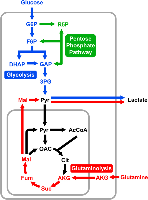
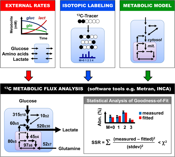
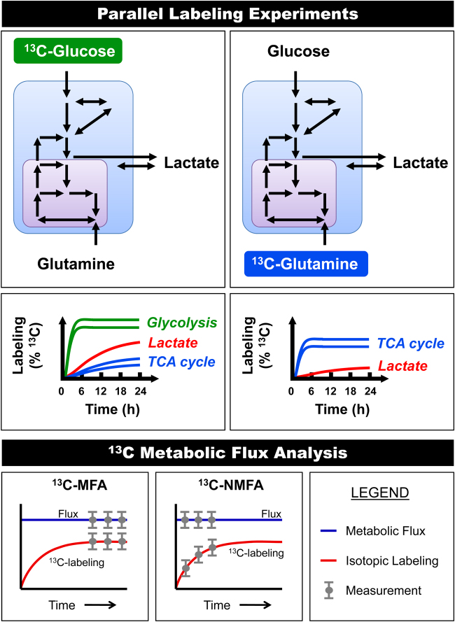
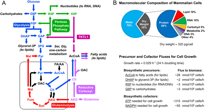
Cancer metabolism is significantly altered from normal cellular metabolism allowing cancer cells to adapt to changing microenvironments and maintain high rates of proliferation. In the past decade, stable-isotope tracing and network analysis have become powerful tools for uncovering metabolic pathways that are differentially activated in cancer cells. In particular, 13C metabolic flux analysis (13C-MFA) has emerged as the primary technique for quantifying intracellular fluxes in cancer cells. In this review, we provide a practical guide for investigators interested in getting started with 13C-MFA. We describe best practices in 13C-MFA, highlight potential pitfalls and alternative approaches, and conclude with new developments that can further enhance our understanding of cancer metabolism.


Conflict of interest statement

The authors declare that they have no conflict of interest.

Figures

Fig. 1
Fig. 1. Glucose and glutamine are the two most highly consumed carbon substrates in cancer cells.
Both substrates can be converted to lactate via glycolysis and glutaminolysis, respectively. High lactate secretion, especially from glucose, is a major hallmark of cancer cells known as the Warburg effect, or aerobic glycolysis
Fig. 2
Fig. 2. 13C metabolic flux analysis (13C-MFA) is a powerful approach for quantifying intracellular metabolic fluxes in cancer cells.
The three inputs required for 13C-MFA are external uptake and secretion rates, isotopic labeling measurements, and a comprehensive compartmentalized model of cellular metabolism. User-friendly software tools for 13C-MFA, such as Metran and INCA, can be used to perform 13C-MFA calculations. These tools produce as outputs fluxes for all reactions in the model, confidence intervals for the estimated fluxes, and statistical analysis of the goodness-of-fit
Fig. 3
Fig. 3. Parallel labeling experiments with different 13C-labeled substrates greatly enhance the resolution of metabolic fluxes in complex models.
The rate of labeling incorporation after the introduction of a 13C-tracer depends on the turnover rate of intracellular metabolites and exchanges between intracellular and extracellular metabolites. In particular, external lactate can slow down labeling of intracellular pyruvate and TCA cycle metabolites from 13C-glucose tracers. If isotopic steady state is reached then labeling data can be analyzed with 13C-MFA. However, if the system has not reached isotopic steady state, then the labeling data must be analyzed using isotopic non-stationary 13C-MFA (13C-NMFA)
Fig. 4
Fig. 4. 13C metabolic fluxes are estimated based on comprehensive compartmentalized models of cellular metabolism.
a The diagram shows important metabolic pathways in cancer metabolism, including glycolysis, pentose phosphate pathway, TCA cycle, reductive carboxylation of glutamine, and transketolase-like 1 (TKTL1) pathway. One of the key functions of cellular metabolism is to supply anabolic building blocks needed for cell growth, shown here as draining reactions from central metabolic pathways. b A typical macromolecular composition of cancer cells is shown. The macromolecular composition and the growth rate of cells determine the rates at which anabolic precursors must be produced to sustain cell growth. Typical values of anabolic precursor fluxes in proliferating cancer cells are shown
Fig. 5
Fig. 5. The isotopomer spectral analysis (ISA) approach is used to quantify de novo fatty acid biosynthesis based on tracer experiments with fully 13C-labeled substrates.
In the classical ISA formulation, two model parameters are determined, the D-value and the g(t)-value. The ISA approach can be generalized and extended to include additional model parameters such as fM2
Fig. 6
Fig. 6. Two alternative 13C-glucose-tracing strategies for analysis of metabolic fluxes in upper metabolism based on mass isotopomer measurements of 3-phosphoglycerate (3PG).
a The [1,2-13C]glucose tracer allows good resolution of relative glycolysis and pentose phosphate pathway fluxes. b A mixture of 50% [2-13C]glucose and 50% [4,5,6-13C]glucose is an improved tracer approach that also allows precise quantification of the transketolase-like 1 (TKTL1) pathway flux
Fig. 7
Fig. 7. [U-13C]Glutamine tracer experiments produce rich labeling patterns in TCA cycle metabolites that allow precise quantification of metabolic fluxes in lower part of central metabolism, i.e., downstream of pyruvate, using 13C-MFA.
The diagram shows schematically the flow of 13C-labeling from [U-13C]glutamine into relevant metabolic pathways in cancer cells. The insert shows an example of labeling data obtained from a [U-13C]glutamine tracer experiment. Colors of arrows indicate different metabolic pathways: reductive carboxylation of glutamine (purple); glutaminolysis (red); conversion of malate to oxaloacetate via malic enzyme and pyruvate carboxylase (green)

References

    1. DeBerardinis RJ, Thompson CB. Cellular metabolism and disease: what do metabolic outliers teach us? Cell. 2012;148:1132–1144. doi: 10.1016/j.cell.2012.02.032. - DOI - PMC - PubMed
    1. Metallo CM, Vander Heiden MG. Understanding metabolic regulation and its influence on cell physiology. Mol. Cell. 2013;49:388–398. doi: 10.1016/j.molcel.2013.01.018. - DOI - PMC - PubMed
    1. Dong W, Keibler MA, Stephanopoulos G. Review of metabolic pathways activated in cancer cells as determined through isotopic labeling and network analysis. Metab. Eng. 2017;43:113–124. doi: 10.1016/j.ymben.2017.02.002. - DOI - PMC - PubMed
    1. Badur MG, Metallo CM. Reverse engineering the cancer metabolic network using flux analysis to understand drivers of human disease. Metab. Eng. 2017;45:95–108. doi: 10.1016/j.ymben.2017.11.013. - DOI - PMC - PubMed
    1. Metallo CM, Deberardinis RJ. Engineering approaches to study cancer metabolism. Metab. Eng. 2017;43:93. doi: 10.1016/j.ymben.2017.09.002. - DOI - PubMed

Publication types

LinkOut - more resources