. 2018 Jan 31;15(1):138-142.
doi: 10.1016/j.jor.2018.01.028. eCollection 2018 Mar.

Bizarre parosteal osteochondromatous proliferation: 16 Cases with a focus on histologic variability

Affiliations

Bizarre parosteal osteochondromatous proliferation: 16 Cases with a focus on histologic variability

Margaret Cocks et al. J Orthop. .

Abstract

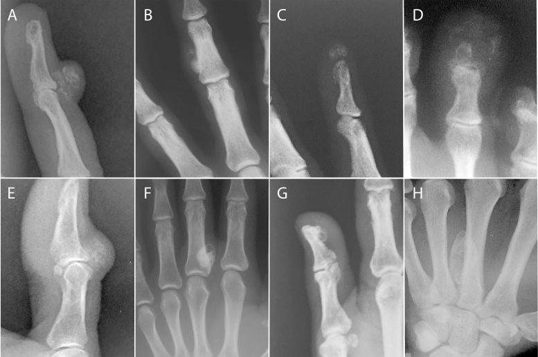
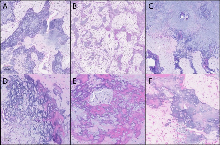
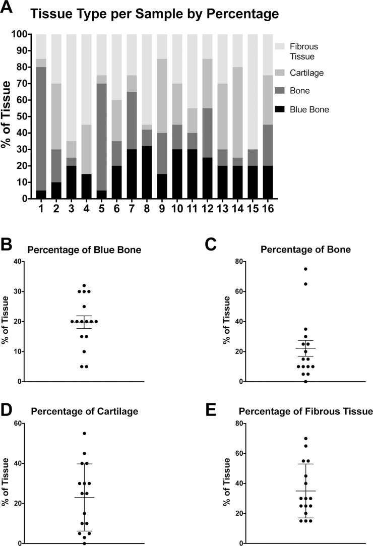
Bizarre parosteal osteochondromatous proliferation (BPOP) is a benign bone and cartilage forming tumor occurring on the surface of bones, predominantly on the hands and feet. A defining feature of BPOP is the purplish-blue mineralization of cartilaginous tissue, known as 'blue bone.' Here, we report on an institutional series of 16 cases of BPOP, including radiographic, histologic, and histomorphometric features. All tumors were composed of some element of bone, cartilage, fibrous tissue and 'blue bone,' though the amount of each tissue sub-type varied widely. Some cases showed focal 'blue bone' only, however this was a defining feature in all cases.

Keywords: BPOP; Bone tumor; Cartilage tumor; Osteochondroma; Surface bone tumor.


Figures

Fig. 1
Fig. 1
Representative radiographic images of bizarre parosteal osteochondromatous proliferation. All were taken of the hands except for D, which is of the toes. (A) Dorsal multilobular radiodensity of the fourth distal phalanx with surrounding soft tissue prominence that has no apparent communication to the underlying bone marrow. (B) Parosteal radiodense mass adjacent to the middle phalanx of the third digit, appears to be a surface lesion. (C) Soft tissue mass with radiodensity distal to the distal phalanx. (D) Soft tissue mass with peripheral radiodensity that is associated with the distal phalanx, probably with periosteal reaction. Appearance of this lesion is less mature than in the prior examples. (E) Dorsal soft tissue lesion with internal radiodensity at the level of the interphalangeal joint of the thumb. Note similarity to radiograph in part A. (F) Radiodense mass at the proximal aspect of the proximal phalanx of the digit. From this view, possible attachment to the adjacent bone cortex cannot be excluded. (G) Volar surface lesion along the distal phalanx with peripheral radiodensity. (H) Large radiodense mass with smooth external border along the proximal metacarpal of the long finger with unclear relationship to the underlying bone.
Fig. 2
Fig. 2
Representative histologic images of bizarre parosteal osteochondromatous proliferation. H&E staining was used on tissue samples. AC were obtained at 40× magnification; DF were obtained at 100× magnification. (A) Representative image of BPOP with abundant ‘blue bone’. (B) Representative image of BPOP with prominent fibrous tissue. (C) Representative images of BPOP with prominent cartilage. In all cases a mixture of fibrous, osseous, and cartilaginous tissue is seen. (DF) At high magnification, a variable appearance of ‘blue bone’ is seen between samples.
Fig. 3
Fig. 3
Histomorphometric analysis of tissue composition among samples of bizarre parosteal osteochondromatous proliferation. Tissue makeup was assessed among various histologic images of BPOP, taken from each of the 16 cases included in the study. Different tissue types were established and their relative amounts were measured in each image as a percentage. A shows this composition for each sample. Each bar numbered 1–16 is a distinct sample corresponding to the numbered samples in Supplemental Table 1. BE show the distribution of percentages in each 40× field for a given tissue type: blue bone, bone, cartilage, and fibrous tissue, respectively. Each dot represents one sample, and the horizontal lines represent the mean and standard error (SEM). (For interpretation of the references to colour in this figure legend, the reader is referred to the web version of this article.)
None

References

    1. Meneses M.F., Unni K.K., Swee R.G. Bizarre parosteal osteochondromatous proliferation of bone (Nora’s lesion) Am J Surg Pathol. 1993;17(7):691–697. - PubMed
    1. Yuen M., Friedman L., Orr W., Cockshott W.P. Proliferative periosteal processes of phalanges: a unitary hypothesis. Skeletal Radiol. 1992;21(5):301–303. - PubMed
    1. Rybak L.D., Abramovici L., Kenan S., Posner M.A., Bonar F., Steiner G.C. Cortico-medullary continuity in bizarre parosteal osteochondromatous proliferation mimicking osteochondroma on imaging. Skeletal Radiol. 2007;36(9):829–834. - PubMed
    1. Abramovici L., Steiner G.C. Bizarre parosteal osteochondromatous proliferation (Nora’s lesion): a retrospective study of 12 cases, 2 arising in long bones. Human Pathol. 2002;33(12):1205–1210. - PubMed
    1. Doganavsargil B., Argin M., Sezak M., Kececi B., Pehlivanoglu B., Oztop F. A bizarre parosteal osteochondromatous proliferation (Nora’s lesion) of metatarsus, a histopathological and etiological puzzlement. Joint Bone Spine. 2014;81(6):537–540. - PubMed