. 2017 Apr 11;27(14):1606243.
doi: 10.1002/adfm.201606243. Epub 2017 Feb 28.

Fluorescent Nanomaterials for the Development of Latent Fingerprints in Forensic Sciences

Affiliations

Fluorescent Nanomaterials for the Development of Latent Fingerprints in Forensic Sciences

Meng Wang et al. Adv Funct Mater. .

Abstract

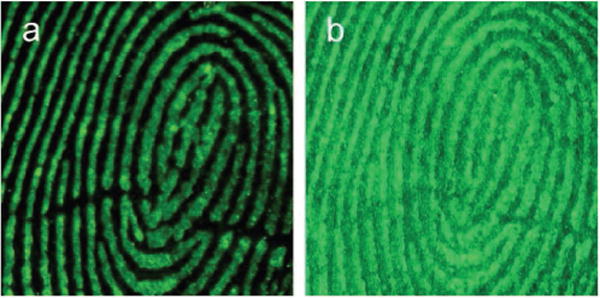
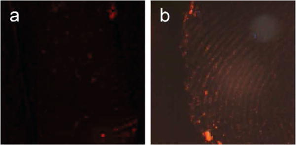
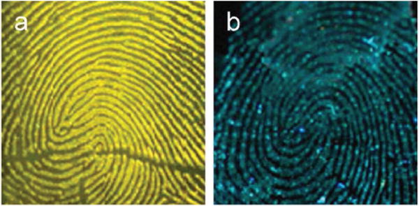
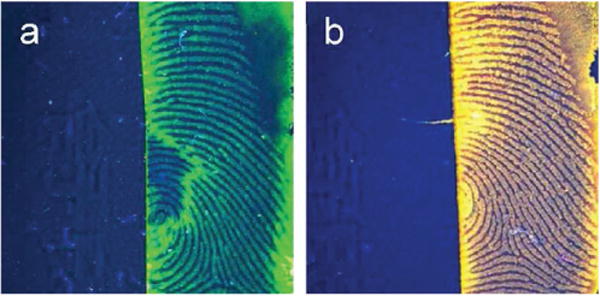
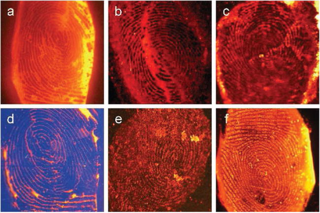
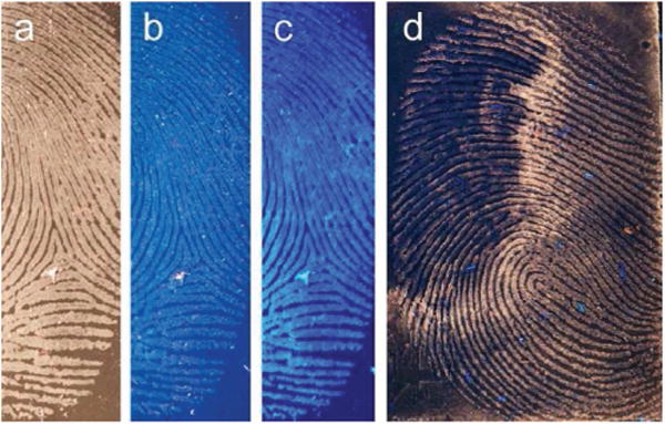
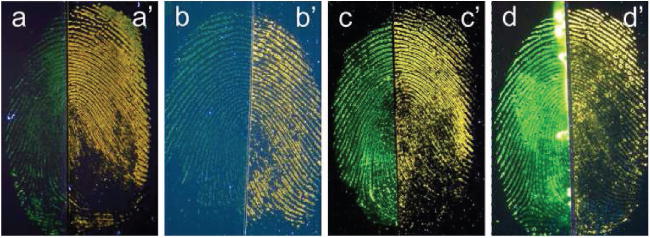
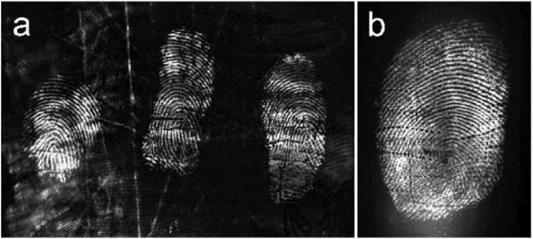
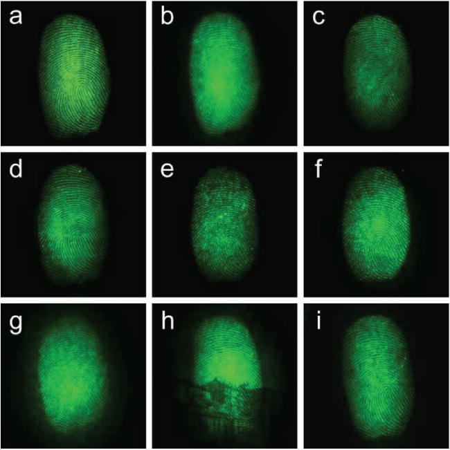
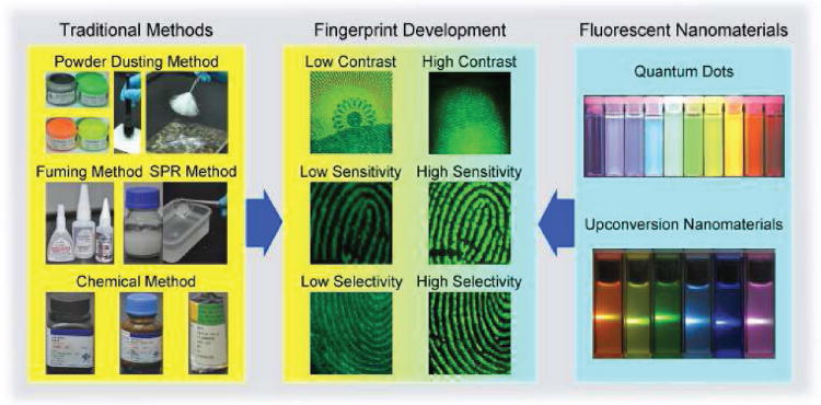
This review presents an overview on the application of latent fingerprint development techniques in forensic sciences. At present, traditional developing methods such as powder dusting, cyanoacrylate fuming, chemical method, and small particle reagent method, have all been gradually compromised given their emerging drawbacks such as low contrast, sensitivity, and selectivity, as well as high toxicity. Recently, much attention has been paid to the use of fluorescent nanomaterials including quantum dots (QDs) and rare earth upconversion fluorescent nanomaterials (UCNMs) due to their unique optical and chemical properties. Thus, this review lays emphasis on latent fingerprint development based on QDs and UCNMs. Compared to latent fingerprint development by traditional methods, the new methods using fluorescent nanomaterials can achieve high contrast, sensitivity, and selectivity while showing reduced toxicity. Overall, this review provides a systematic overview on such methods.


Figures

Figure 1
Figure 1
Latent fingerprint development on Chinese paper money through the fluorescent property of different developing powders: a) NaYF4:Yb,Er UCNMs, in dark field and under 980 nm NIR excitation; and (b) green fluorescent powders, in dark field and under 254 nm UV excitation. Reproduced with permission.[74] Copyright 2015, American Chemical Society.
Figure 2
Figure 2
Latent fingerprint development on glass by using different types of developing powders: a) NaYF4:Yb, Er UCNMs, in dark field and under 980 nm NIR excitation; and (b) bronze flake, in bright field. Reproduced with permission.[73] Copyright 2014, Springer.
Figure 3
Figure 3
Latent fingerprint development on glass by using different types of developing powders: a) NaYF4:Yb, Er UCNMs, in dark field and under 980 nm NIR excitation; and (b) green fluorescent powders, in dark field and under 254 nm UV excitation.
Figure 4
Figure 4
Aqueous suspensions of CdSe@ZnS QDs with ten distinguishable fluorescent emission colors excited with a 350 nm UV radiation. From left to right, the maximum emissions are located at 443, 473, 481, 500, 518, 543, 565, 587, 610, and 655 nm. Reproduced with permission.[47] Copyright 2001, Nature Publishing Group.
Figure 5
Figure 5
Latent fingerprint development on aluminum by using CdS/chitosan NCs under the 450 nm light excitation and imaged with a) 550 nm long pass barrier filter and b) 565 nm band pass barrier filter. Reproduced with permission.[53] Copyright 2009, Elsevier.
Figure 6
Figure 6
Latent fingerprint development by using CdS/PPH NCs under the 450 nm light excitation on the different substrates: a) steel tweezers, and b) glass. Reproduced with permission.[54] Copyright 2011, Elsevier.
Figure 7
Figure 7
Latent fingerprint development by using CdTe/MMT NCs under the 365 nm UV excitation on a variety of substrates: a) polymer, b) painted wood, c) glass, and (d) leather. Reproduced with permission.[35] Copyright 2011, American Chemical Society.
Figure 8
Figure 8
Latent fingerprint development by using CdTe@SiO2 NMs under the 365 nm UV excitation on a variety of substrates: a) glass, b) polymer materials, c) aluminum foil, d) black ceramic, e) black rubber, and f) paper. Reproduced with permission.[55] Copyright 2012, Springer.
Figure 9
Figure 9
Latent fingerprint development by using CdS/DSS NCs under the UV excitation on different substrates: a) aluminum foil, and b) soft drink can. Reproduced with permission.[56] Copyright 2000, ASTM International.
Figure 10
Figure 10
Latent fingerprint development on the aluminum foils by using CdS/PAMAM NCs dissolved in a) methanol, and b) 1:9 methanol-water solutions, under the 365 nm UV light excitation with the assistance of a yellow light filter and a blue light filter, respectively. Reproduced with permission.[57] Copyright 2008, Elsevier.
Figure 11
Figure 11
Latent fingerprint development on the sticky side of tapes by using a) CdSe and b) CdSe@CdS QDs under the 380 nm UV excitation. Reproduced with permission.[58] Copyright 2009, Elsevier.
Figure 12
Figure 12
Latent fingerprint development on the sticky side of adhesives by using CdTe QDs with multi-colors under the 365 nm UV excitation: a) CdTe QDs with green fluorescence, synthesized without refluxing, b) CdTe QDs with yellow fluorescence, refluxed for 2 h. Reproduced with permission.[37] Copyright 2010, Elsevier.
Figure 13
Figure 13
Latent fingerprint development by using positively charged CdTe QDs under the 365 nm UV excitation on a variety of substrates: a) glass, b) black ceramic, c) painted polymer material, d) transparent plastic sheet, e) rough plastic sheet, and f) black rubber. Reproduced with permission.[36] Copyright 2011, IOP Publishing.
Figure 14
Figure 14
Latent fingerprint development on the sticky side of black electrical tape by using CdSe QDs: a) in the bright field, b, d) in the dark field with 365 nm UV excitation, and c) in the dark field with 440 nm blue light excitation. Reproduced with permission.[59] Copyright 2014, Elsevier.
Figure 15
Figure 15
Blood fingerprint development by using CdTe QDs (left halves, a–d) and AY7 (right halves, a′–d′) under the 300–400 nm UV excitation on a variety of substrates: (a, a′) glass, (b, b′) transparent polypropylene, (c, c′) black polyethylene, and (d, d′) aluminum. Reproduced with permission.[61] Copyright 2009, Elsevier.
Figure 16
Figure 16
Blood fingerprint development by using ZnS:Cu QDs (left haves, a–d; right halves, e′–h′), AY7 (right halves, a′–d′), and CdTe QDs (left halves, e–h), under the 300–400 nm UV excitation on a variety of substrates: (a, a′, e, e′) glass, (b, b′, f, f′) transparent polypropylene, (c, c′, g, g′) lack polyethylene, and (d, d′, h, h′) aluminum. Reproduced with permission.[62] Copyright 2013, Elsevier.
Figure 17
Figure 17
Aqueous suspensions ofRE3+ ions-doped NaYbF4 UCNMs with six distinguishable fluorescent emission colors excited with a 980 nm NIR radiation. From left to right, the UCNMs are NaYbF4:Er, NaYbF4:Er,Ho, NaYbF4:Ho, NaYbF4:Tm,Ho, NaYbF4:Tm, and NaYbF4:Er,Tm. Reproduced with permission.[65] Copyright 2009, American Chemical Society.
Figure 18
Figure 18
Latent fingerprint development on Australian five-dollar polymer banknote by using a) NaYF4:Yb,Er UC powders and b) YVO4:Yb,Er UC powders under the 980 nm NIR excitation. (a) Reproduced with permission[71] Copyright 2011, Elsevier. (b) Reproduced with permission.[72] Copyright 2012, Elsevier.
Figure 19
Figure 19
Latent fingerprint development by using NaYF4:Yb,Er UCNMs under the 980 nm NIR excitation on a variety of substrates: a) aluminum alloys sheets, b) stainless steel sheets, c) aluminum foils, d–e) plastic cards, f) floor leathers, g) ceramic tiles, h) wood floor, and i) painted wood. Reproduced with permission.[73] Copyright 2014, Springer.
Figure 20
Figure 20
Latent fingerprint development by using NaYF4:Yb,Er UCNMs under the 980 nm NIR excitation. a–c) On the substrates with a single background color: (a) glass, (b) white ceramic tiles, and (c) black marbles. d–f) On the substrates with background color distraction: various marbles with different surface textures. g–i) On the substrates with background fluorescence interference: (g) note papers, (h) Chinese paper money, and (i) fluorescent plastic plates. Reproduced with permission.[74] Copyright 2015, American Chemical Society.
Figure 21
Figure 21
a) Image of the marble with three latent fingerprints in the black circles, and the developers from top to bottom are FAM/LBA, CdTe/LBA, and NaYF4:Yb,Er/LBA, respectively. b–d) Images of fingerprints treated by (b) FAM, (c) CdTe QDs, and (d) NaYF4:Yb,Er UCNMs-based developers: from top to bottom, the images in row 1 are fingerprints treated by FAM, CdTe QDs, and NaYF4:Yb,Er UCNMs, respectively; the images in row 2 are fingerprints treated by FAM/LBA, CdTe/LBA, and NaYF4:Yb,Er/LBA, respectively; the images in row 3 are the corresponding magnified images of row 2. Reproduced with permission[38] Copyright 2014, John Wiley and Sons.
Figure 22
Figure 22
Development of latent, fresh (2 h old) fingerprints by using a NaYF4:Yb,Er UCNMs based suspension on a variety of substrates: a) stainless steel sheets, b) aluminum alloys sheets, c) aluminum foils, d) marbles, e) ceramic tiles, f) plastic cards, g) painted wood, and h) Chinese paper money. Development of latent, aged (1-year-old) fingerprints by using NaYF4:Yb,Er UCNMs-based suspension on glass is shown in i). The left panels (except that in (h)) are images in a bright field without 980 nm irradiation; the left panel of (h) is the image under 254 nm UV excitation. Right panels show fluorescent images formed under 980 nm NIR excitation. Reproduced with permission.[32] Copyright 2016, Royal Society of Chemistry.
Scheme 1
Scheme 1
General idea of this review. Upper image in the right panel: Reproduced with permission.[47] Copyright 2001, Nature Publishing Group. Lower image at right panel: Reproduced with permission.[65] Copyright 2009, American Chemical Society. Images of the fingerprints in the middle pannel: Reproduced with permission.[74] Copyright 2015, American Chemical Society.

References

    1. Saferstein R. Criminalistics: An Introduction to Forensic Science. 9th. Prentice Hall; Englewood Cliffs, NJ: 2006.
    1. Jackson ARW, Jackson JM. Forensic Science. 2nd. Prentice Hall; Harlow, England: 2008.
    1. Champod C, Lennard C, Margot P, Stoilovic M. Fingerprints and Other Ridges Skin Impressions. CRC Press; Boca Raton, FL: 2004.
    1. Maltoni D, Maio D, Jain A, Prabhakar S. Handbook of Fingerprint Recognition, Springer-Verlag, New York. 2009
    1. Faulds H. Nature. 1880;22:605.

LinkOut - more resources