

Imaging Characteristics of Dural Arteriovenous Fistulas Involving the Vein of Galen: A Comprehensive Review
- PMID: 29657906
- PMCID: PMC5896872
- DOI: 10.7759/cureus.2180
Imaging Characteristics of Dural Arteriovenous Fistulas Involving the Vein of Galen: A Comprehensive Review
Abstract
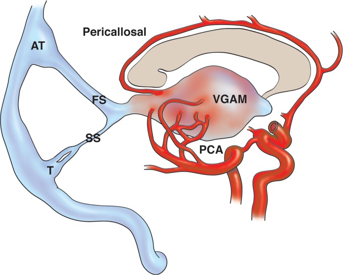
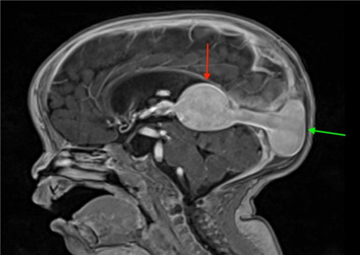
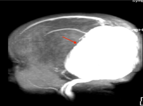
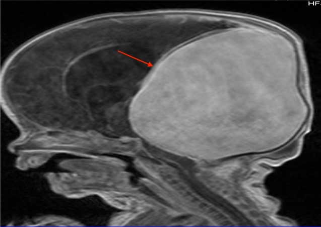
Vein of Galen aneurysmal malformation (VGAM) is a rare angiopathy, which most commonly presents in infancy. Although very rare, it is associated with high morbidity and mortality rates. In order to minimize such morbid rates, a prompt diagnosis followed by a timely initiation of management is crucial. Multiple antenatal and postnatal imaging techniques for the diagnosis have been described and discussed in the literature. However, to our knowledge, a comprehensive review exploring such a list of imaging options for VGAM has never been established. We aim to review the diagnostic tools to aid in better understanding of the investigative modalities physicians may choose from when treating patients with a VGAM.
Keywords: aneurysmal malformation; arteriovenous fistula; diagnosis; imaging; prenatal; vein of galen.
Conflict of interest statement
The authors have declared that no competing interests exist.
Figures
References
-
- CT and color Doppler diagnosis of the vein of Galen malformations with hydrocephalus: a case series report of rare intracranial vascular malformations. Kebede T, Hawaz Y, Assefa G. http://europepmc.org/abstract/med/23930495. Ethiop Med J. 2013;51:77–83. - PubMed
-
- Prenatal diagnosis of a vein of Galen aneurysmal malformation using color Doppler ultrasound: a case report. Lopez-Cepero R, de la Vega A, Lynch L. http://europepmc.org/abstract/med/23767382. Bol Asoc Med P R. 2013;105:32–35. - PubMed
-
- Vein of Galen aneurysmal malformation-clinical and angiographic spectrum with management perspective: an institutional experience. Agarwal H, Sebastian LJ, Gaikwad SB, Garg A, Mishra NK. J Neurointerv Surg. 2017;9:159–164. - PubMed
-
- Galenic pial arteriovenous fistulas: Angioarchitecture, clinical presentation, and therapeutic considerations. George Zaki Ghali M. Clin Anat. 2017 - PubMed
Publication types
LinkOut - more resources
Full Text Sources
Other Literature Sources