

Distribution of Thoracic Aortic Calcifications in Patients Undergoing Coronary Artery Bypass Grafting
- PMID: 29657951
- PMCID: PMC5890763
- DOI: 10.12945/j.aorta.2017.17.035
Distribution of Thoracic Aortic Calcifications in Patients Undergoing Coronary Artery Bypass Grafting
Abstract
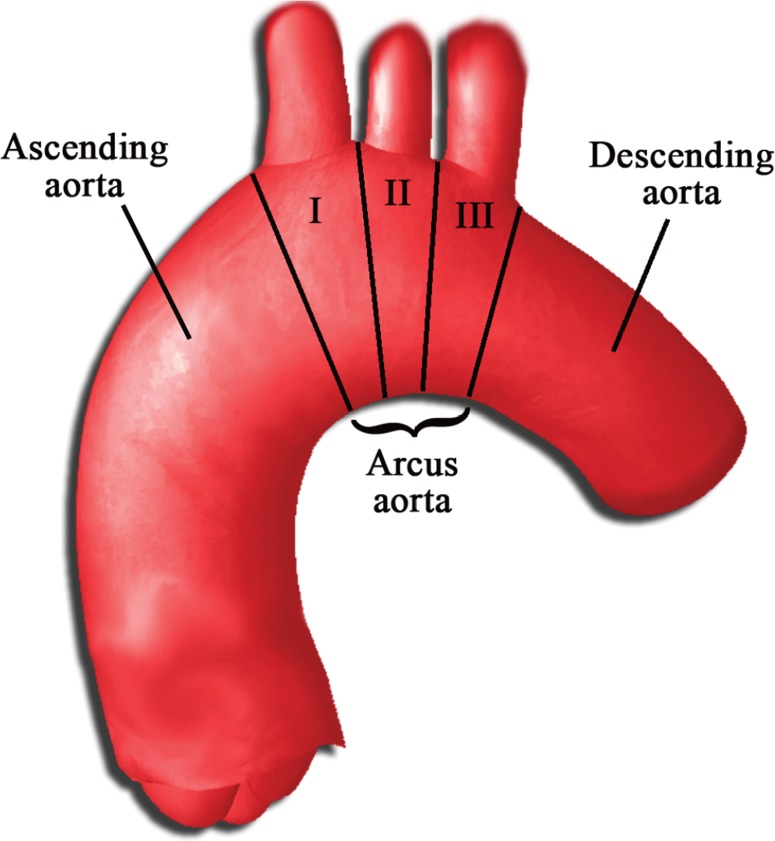
Background: In procedures involving surgical maneuvers such as cannulation, clamping, or proximal anastomosis where aortic manipulation is inevitable, a preliminary assessment of atherosclerotic plaques bears clinical significance. In the present study, our aim was to evaluate the frequency and distribution of aortic calcifications in patients undergoing coronary artery bypass grafting (CABG) surgery to propose a morphological classification system.
Methods: A total of 443 consecutive patients with coronary artery disease were included in this study. Preoperative non-contrast enhanced computed tomography images, in-hospital follow-up data, and patient characteristics were retrospectively evaluated.
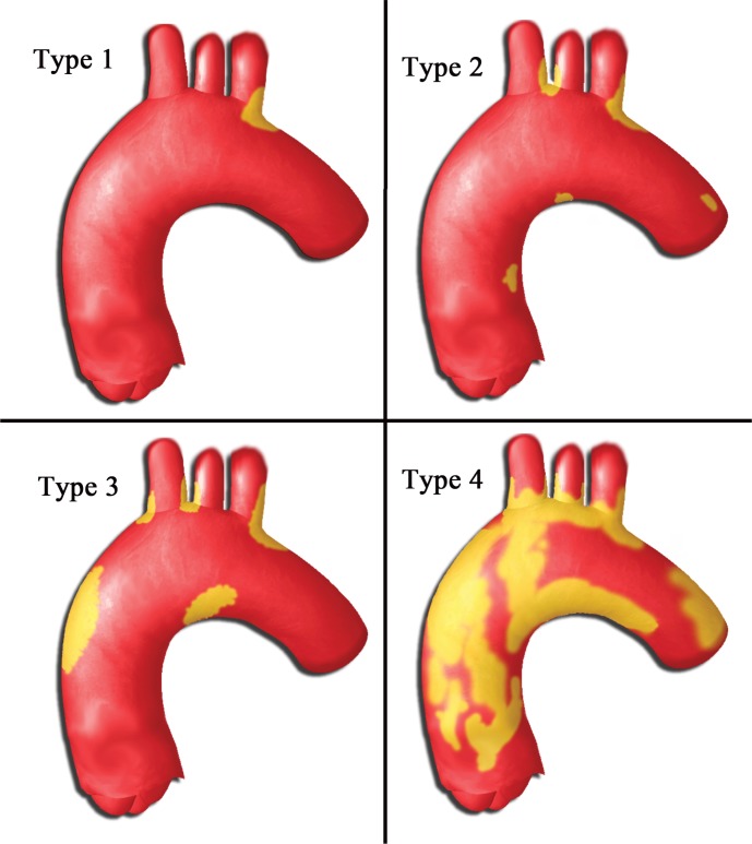
Results: Whereas 33% of patients had no calcifications at any site in the aorta, 7.9%, 75.4%, and 16.7% had calcifications in the ascending aorta, aortic arch, and descending aorta, respectively. Focal small calcifications were the most common type of lesions in the ascending aorta (3.9%), whereas 9 patients (1.4%) had porcelain ascending aorta. We defined four types of patients with increasing severity and extent of calcifications.
Conclusions: Based on the frequency and distribution of calcifications in the thoracic aorta, we propose a classification system from least to most severe for coronary artery disease patients who are candidates for CABG.
Keywords: Calcification; Coronary artery bypass; grafting Aorta.
Figures
References
LinkOut - more resources
Full Text Sources
Other Literature Sources