. 2017;1(1):7-18.

Method for the Culture of Mouse Pulmonary Microvascular Endothelial Cells

Affiliations

Method for the Culture of Mouse Pulmonary Microvascular Endothelial Cells

Anita Kovacs-Kàsa et al. Sci Pages Pulmonol. 2017.

Abstract

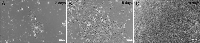
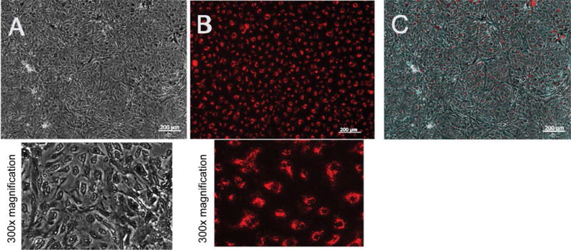
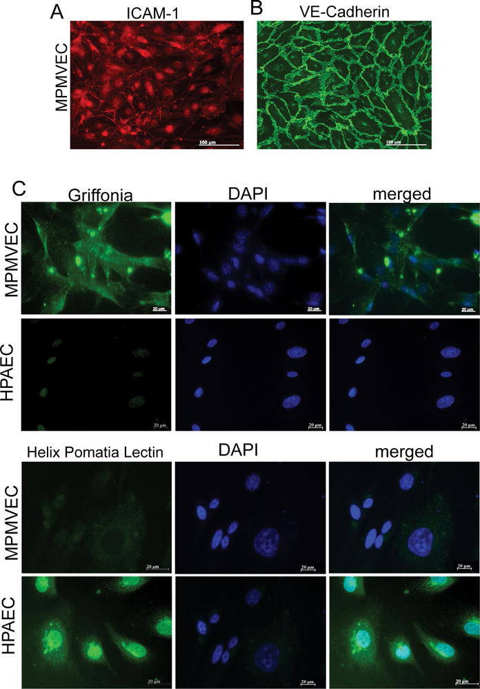
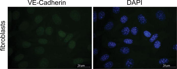
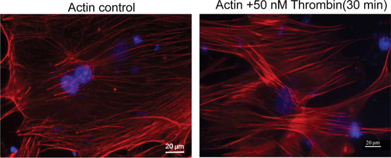
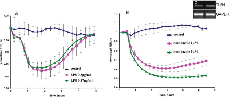
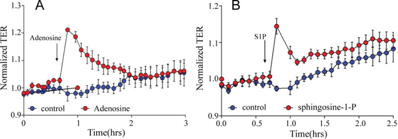
Pulmonary microvascular endothelial cells (ECs) are integral to the alveoli-capillary barrier of the lung. The EC barrier integrity is known to be disrupted in severe lung diseases such as acute respiratory distress syndrome (ARDS), pneumonia and pulmonary edema. Mice are commonly used to model these diseases, dictating an increasingly high demand for murine ECs isolation and culture. Despite the significant number of protocols for the culture of various types of murine cells, the isolation of microvascular endothelial cells remains a challenging procedure. In our manuscript we developed adetailed step-by-step refined method for isolation murine pulmonary microvascular ECs for in vitro studies. We separated cells using platelet endothelial cell adhesion molecule antibody and characterized ECs with antibodies against intercellular adhesion molecule-1, acetylated-low density lipoprotein, and vascular endothelial (VE)-cadherin. Further, we confirmed microvascular origin of these cells using Griffonia simplicifolia and Helix pomatia (negative control) staining. Barrier properties of EC monolayer were characterized by conducting electric cell-substrate impedance sensing experiments with the edemagenic agents, lipopolysaccharide and nocodazole, and known barrier-protective agents, adenosine and sphingosine-1-phosphate. The described complete protocol provided consistent and reproducible results.

Keywords: Cell culture; Lung; Method; Microvascular endothelial cells; Murine pulmonary endothelial cells.


Conflict of interest statement

Disclosure/Duality of Interest No author has any disclosure or duality of interest for any product or result in this manuscript.

Figures

Figure 1
Figure 1
Nesting of mouse pulmonary microvascular ECs. Phase contrast micrographs of mouse pulmonary microvascular endothelial cells culture on (A) day-2 showing Dynabeads® in culture and an inset with a “nest” of endothelial cells and; (B) day-6; (C) Shows a phase-contrast confluent monolayer on day-8. Scale bars are 200 μm.
Figure 2
Figure 2
Labeling of mouse pulmonary microvascular endothelial cells with Ac-LDL. Labeling of mouse pulmonary microvascular endothelial cellson day-6 (passage 2) with acetylated-low density lipoprotein cultured at 37 °C in complete endothelial growth basal medium-2 (Lonza, Allendale, New Jersey, USA) on fibronectin (A) Phase-contrast micrograph of a confluent monolayer and below is 300× magnifications; (B) Uptake of acetylated-low density lipoprotein and below is 300× magnification; (C) Confluent monolayer merged with acetylated-low density lipoprotein. Scale bars are 200 μm.
Figure 3
Figure 3
A Characteristics of mouse pulmonary microvascular endothelial cells (MPMVECs). MPMVECs were cultured at 37 °C in complete endothelial growth basal medium-2 on fibronectin until confluent (A) Expression of Intercellular adhesion molecule-1 (ICAM-1) in representative micrograph of cultured endothelial cells; (B) Expression of VE-cadherin is localized at the cell-cell junctions shown in representative micrograph with expression of vascular endothelial cadherin; (C) Endothelial cells isolated from murine lungs or HPAEC stained either with Griffonia or Helix Pomatia lectin. Scale bars are 20 μm.
Figure 4
Figure 4
NIH3T3 fibroblasts are stained with DAPI and VE-cadherin. Fibroblasts were stained with cultured NIH3T3 cells grown on glass coverslips. Cells were fixed in a 3.7% formaldehyde solution in PBS for 10 min at and washed three times with PBS. The cells were permeabilized with 0.2% Triton X-100 in TBS supplemented with 0.1% Tween 20 (TBST) for 5 min, washed three times with PBS and blocked with 2% BSA in TBST for 1 h. Incubation with specific antibodies diluted with a blocking solution was performed for 1 h at room temperature. Specific antibody was used to detect VE-cadherin. After three washes with PBS, the cells were incubated with appropriate secondary antibody (1:300) conjugated with fluorescent dye Alexa 488 (green) 1 h at room temperature. The coverslips were mounted with proLong antifade reagent with DAPI. After immunostaining, the cells were analyzed using Zeiss Axiolab microscope using 63 × oil immersion objective. Scale bars are 20 μm.
Figure 5
Figure 5
Thrombin treatment induced increased actin cytoskeleton rearrangement in MPMVEC. MPMVECs were cultured at 37 °C in complete endothelial growth basal medium-2 until confluent. The cells grown on coverslips and fixed with 3.7% formaldehyde solution in PBS for 10 min and washed 3 times with PBS and stained with Texas Red®-phalloidin for actin. Untreated cells are compared to thrombin treatment (50 nM) × 30 min. Visualization was performed using microscope (Axiolab; Carl Zeiss, Oberkochen, Germany) at 63 × oil immersion objective. Scale bars are 20 μm.
Figure 6
Figure 6
Time-dependent decrease in trans-endothelial resistance (TER) of mouse pulmonary microvascular endothelial cells (A) LPS (0.3 μg/mL or 0.17 μg/mL) and (B) nocodazole (1 μM or 5 μM). Eight-well arrays were inoculated with mouse pulmonary microvascular endothelial cells (50,000); 48–72 h later, confluent monolayers were observed exhibiting TER of ≈800 MΩ to ≈900 MΩ. (A) LPS or; (B) Nocodazole or veh were added and TER values recorded continuously at 20-s intervals over the next 6 h. Data are representative of three separate experiments; (C) PCR analysis for TLR4 expression (Inset) RT-Polymerase chain reaction analysis for toll like receptor-4 (TLR4) expression in endothelial cell from murine lungs. GAPDH expression was used as a housekeeping control.
Figure 7
Figure 7
Trans-endothelial resistance (TER) of the barrier protective agents adenosine and Sphingosine-1-phosphate in mouse pulmonary microvascular endothelial cells. Eight-well arrays were inoculated with mouse pulmonary microvascular endothelial cells (50,000); 48–72 h later, confluent monolayers were observed exhibiting TER of ≈800 MΩ to ≈900 MΩ. (A) adenosine orveh; (B) Sphingosine-1-phosphate orveh were added and TER values recorded continuously at 20-s intervals over the next 3 h. Data are representative of three separate experiments.

Similar articles

Cited by

References

    1. Akis N, Madaio MP. Isolation, culture, and characterization of endothelial cells from mouse glomeruli. Kidney Int. 2004;65:2223–2227. - PubMed
    1. Wu Z, Hofman FM, Zlokovic BV. A simple method for isolation and characterization of mouse brain microvascular endothelial cells. J Neurosci Methods. 2003;130:53–63. - PubMed
    1. Matute-Bello G, Frevert CW, Martin TR. Animal models of acute lung injury. Am J Physiol Lung Cell Mol Physiol. 2008;295:379–399. - PMC - PubMed
    1. Marelli-Berg FM, Peek E, Lidington EA, et al. Isolation of endothelial cells from murine tissue. J Immunol Methods. 2000;244:205–215. - PubMed
    1. Czikora I, Sridhar S, Gorshkov B, et al. Protective effect of Growth Hormone-Releasing Hormone agonist in bacterial toxin-induced pulmonary barrier dysfunction. Front Physiol. 2014;5:259. - PMC - PubMed

LinkOut - more resources