Review
. 2018 Nov;91(1091):20180074.
doi: 10.1259/bjr.20180074. Epub 2018 Jun 15.

Scandium and terbium radionuclides for radiotheranostics: current state of development towards clinical application

Affiliations
Review

Scandium and terbium radionuclides for radiotheranostics: current state of development towards clinical application

Cristina Müller et al. Br J Radiol. 2018 Nov.

Abstract

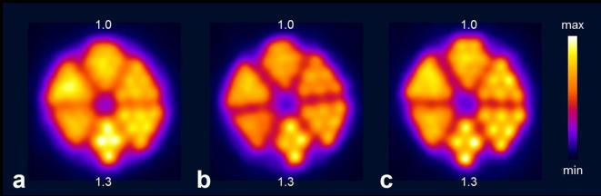
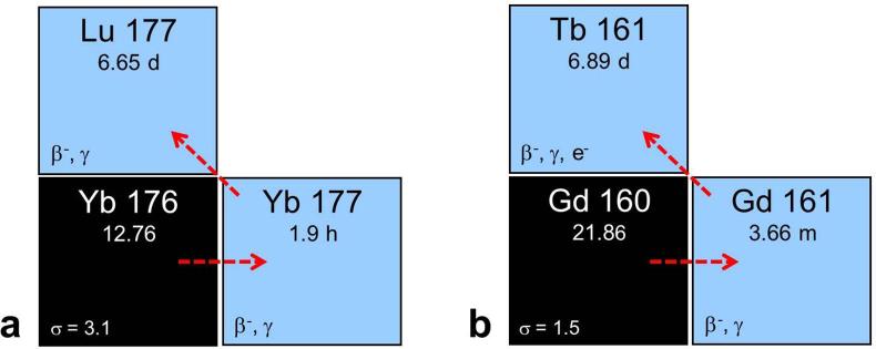
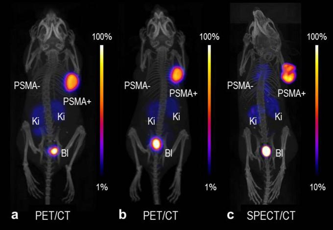
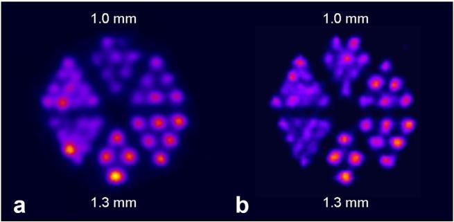
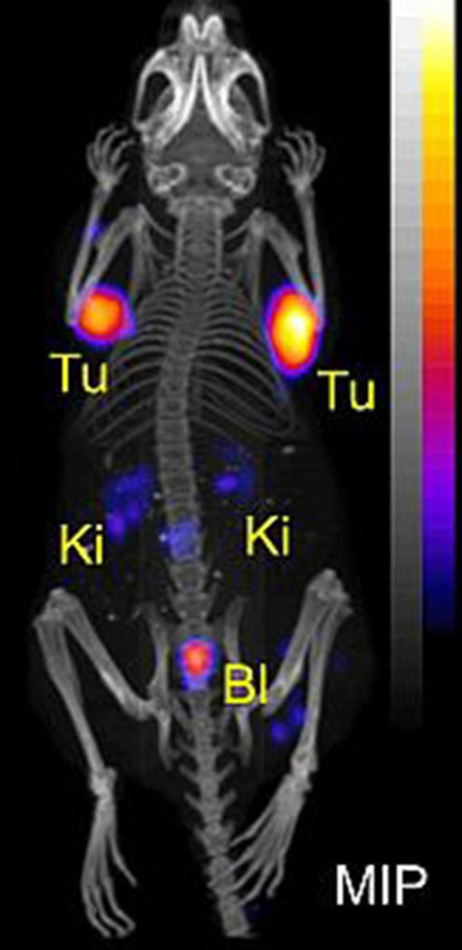
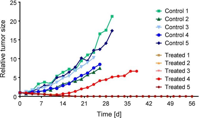
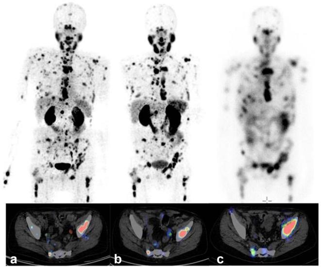
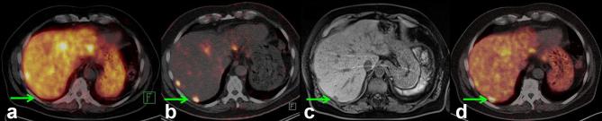
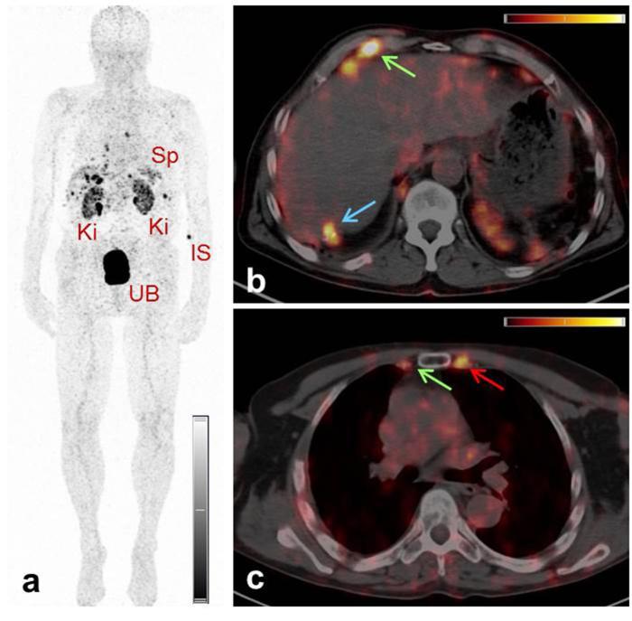
Currently, different radiometals are in use for imaging and therapy in nuclear medicine: 68Ga and 111In are examples of nuclides for positron emission tomography (PET) and single photon emission computed tomography (SPECT), respectively, while 177Lu and 225Ac are used for β-- and α-radionuclide therapy. The application of diagnostic and therapeutic radionuclides of the same element (radioisotopes) would utilize chemically-identical radiopharmaceuticals for imaging and subsequent treatment, thereby enabling the radiotheranostic concept. There are two elements which are of particular interest in this regard: Scandium and Terbium. Scandium presents three radioisotopes for theranostic application. 43Sc (T1/2 = 3.9 h) and 44Sc (T1/2 = 4.0 h) can both be used for PET, while 47Sc (T1/2 = 3.35 d) is the therapeutic match-also suitable for SPECT. Currently, 44Sc is most advanced in terms of production, as well as with pre-clinical investigations, and has already been employed in proof-of-concept studies in patients. Even though the production of 43Sc may be more challenging, it would be advantageous due to the absence of high-energetic γ-ray emission. The development of 47Sc is still in its infancy, however, its therapeutic potential has been demonstrated preclinically. Terbium is unique in that it represents four medically-interesting radioisotopes. 155Tb (T1/2 = 5.32 d) and 152Tb (T1/2 = 17.5 h) can be used for SPECT and PET, respectively. Both radioisotopes were produced and tested preclinically. 152Tb has been the first Tb isotope that was tested (as 152Tb-DOTATOC) in a patient. Both radionuclides may be of interest for dosimetry purposes prior to the application of radiolanthanide therapy. The decay properties of 161Tb (T1/2 = 6.89 d) are similar to 177Lu, but the coemission of Auger electrons make it attractive for a combined β-/Auger electron therapy, which was shown to be effective in preclinical experiments. 149Tb (T1/2 = 4.1 h) has been proposed for targeted α-therapy with the possibility of PET imaging. In terms of production, 161Tb and 155Tb are most promising to be made available at the large quantities suitable for future clinical translation. This review article is dedicated to the production routes, the methods of separating the radioisotopes from the target material, preclinical investigations and clinical proof-of-concept studies of Sc and Tb radionuclides. The availability, challenges of production and first (pre)clinical application, as well as the potential of these novel radionuclides for future application in nuclear medicine, are discussed.


Figures

Figure 1.
Figure 1.
Transversal sections of PET scans of Derenzo phantoms (hole diameter ranging from 0.8 to 1.3 mm in 0.1 mm increments; 1.0 and 1.3 indicate the holes of 1.0 mm and 1.3 mm diameter, respectively) filled with (a) > 99% 44Sc, (b) 66.2% 43Sc/33.3% 44Sc and (c) 98.2% 43Sc. The acquisition of the PET scans was performed within the energy window of 400–700 keV for 30 min, in order to obtain a total number of ~6 × 107 coincidences. Figure adapted from Domnanich et al. EJNMMI Radiopharmacy and Chemistry 2017; 2:14. PET, positron emission tomography.
Figure 2.
Figure 2.
(a) Production route of no-carrier-added 177Lu via the 176Yb(n,γ)177Yb→177Lu nuclear reaction. (b) Analogous production route of 161Tb via the 160Gd(n,γ)161Gd→161Tb nuclear reaction. Figure adapted from Karlsruhe nuclide chart, 8th edition, 2012 (https://www.nucleonica.com/).
Figure 3.
Figure 3.
PET/CT and SPECT/CT images as MIPs of mice 2 h after injection of (a) 43Sc-PSMA-617, (b) 44Sc-PSMA-617 and (c) 47Sc-PSMA-617. PSMA-=PC-3 flu tumor; PSMA+=PC-3 PIP tumor, Ki = kidney, Bl = urinary bladder. The PET/CT and SPECT/CT images were prepared using VivoQuant software. The scales of PET and SPECT images were cut by 1 and 10%, respectively, to make the accumulated activity in tumors and kidneys better visible (unpublished results). BI, urinary bladder; Ki, kidney; MIPs, maximum intensity projections; PET, positron emission tomography; PSMA, prostate-specific membrane antigen; SPECT, single photon emission computed tomography.
Figure 4.
Figure 4.
SPECT images of Derenzo phantoms filled with (a) 155Tb (2.6 MBq) and (b) 111In (4 MBq). Figure adapted from Müller et al. Nucl Med Biol 2014;41 Suppl:e58-65.3] SPECT, single photon emission computed tomography.
Figure 5.
Figure 5.
MIP of PET/CT image of AR42J tumor-bearing mouse 2 h after injection of 149Tb-DOTATOC (7 MBq). Figure adapted from Müller et al. EJNMMI Radiopharmacy and Chemistry 2016;1:5. BI, urinary bladder; Ki, kidney; MIP, maximum intensity projection; PET, positron emission tomography; Tu, tumor.
Figure 6.
Figure 6.
Pre-clinical therapy study: the graph represents the tumor growth relative to the average tumor size (tumor volume) determined at Day 0, which was set to 1 (indicated as relative tumor size). The study was performed with five untreated mice (controls: green/blue) and five mice treated with 161Tb-folate (11 MBq/mouse; red/orange). In the treated group, four of the five mice showed complete tumor remission as shown by overlapping graphs. This research was originally published in JNM, Müller et al. PET, positron emission tomography; SPECT, single photon emission computed tomography.
Figure 7.
Figure 7.
MIP (top) and representative sections (bottom) of PET/CT examination of a patient suffering from mCRPC, with high tumor load, using (a) 44Sc-PSMA-617 (50 MBq, 60 min p.i.), and (b) 68Ga-PSMA-11 (120 MBq, 60 min p.i.). (c) On the right hand side, the planar scintigraphy is shown (top) and a representative section of the post-therapy SPECT/CT scan, about 24 h after application of 6.7 GBq 177Lu-PSMA-617. Figure adapted from Eppard et al. Theranostics 2017;7:4359–69. mCRPC, metastasized castration-resistant prostate cancer; MIP, maximum intensity projection; PET, positron emission tomography; PSMA, prostate-specific membrane antigen.
Figure 8.
Figure 8.
Comparison of serial images of the transverse section of liver, representing the lesion in segment VII (green arrow), obtained by PET/CT imaging of Patient 1 using somatostatin analogs for restaging after the third cycle of PRRT. (a) At 18 months after PRRT, the lesion was not detected on PET/CT image obtained with 68Ga-DOTATOC; (b) at 27 months after PRRT, the lesion was detected using 44Sc-DOTATOC; (c) a concurrent MRI performed within 24 h of the PET/CT scan obtained with 44Sc-DOTATOC co-registered the lesion seen on the PET/CT image; (d) at 36 months after PRRT, the lesion was detected on PET/CT images obtained with 68Ga-DOTATOC. Figure adapted from Singh et al. Cancer Biother Radiopharm 2017;32:124–32 [31] PET, positron emission tomography; PRRT, peptide receptor radionuclide therapy.
Figure 9.
Figure 9.
PET/CT images of a patient with neuroendocrine neoplasm of the terminal ileum obtained at 2 h after injection of 152Tb-DOTATOC. (a) MIP image shows the Ki, the UB, the Sp and the IS. (b/c) Transverse sections of PET/CT fusion images demonstrate radiopeptide uptake in lymph node metastases in the right costophrenic region and in the right internal mammary chain (green arrows), as well as in Segment 7 of the liver (blue arrows) and in a skeletal metastasis in the left third rib adjacent to the sternocostal junction (red arrows). Figure adapted from Baum et al. Dalton Trans 2017;46:14638–46. IS, injection site; Ki, kidneys; MIP, maximum intensity projection; Sp, spleen; UB, urinary bladder.

References

    1. Yordanova A, Eppard E, Kürpig S, Bundschuh RA, Schönberger S, Gonzalez-Carmona M, et al. . Theranostics in nuclear medicine practice. Onco Targets Ther 2017; 10: 4821–8. doi: 10.2147/OTT.S140671 - DOI - PMC - PubMed
    1. Ahn BC. Personalized medicine based on theranostic radioiodine molecular imaging for differentiated thyroid cancer. Biomed Res Int 2016; 2016: 1–9. doi: 10.1155/2016/1680464 - DOI - PMC - PubMed
    1. Hertz S, Roberts A. Radioactive iodine as an indicator in thyroid physiology V. The use of radioactive iodine in the differential diagnosis of two types of Graves' disease. J Clin Invest 1942; 21: 31–2. doi: 10.1172/JCI101276 - DOI - PMC - PubMed
    1. Hertz B, Schuller K, Saul Hertz M. Saul Hertz, MD (1905–1950): A pioneer in the use of radioactive Iodine. Endocr Pract 2010; 16: 713–5. doi: 10.4158/EP10065.CO - DOI - PubMed
    1. Padma S, Sundaram PS. Radioiodine as an adjuvant therapy and its role in follow-up of differentiated thyroid cancer. J Cancer Res Ther 2016; 12: 1109–13. doi: 10.4103/0973-1482.163677 - DOI - PubMed

MeSH terms