Review
. 2018 Apr 16;19(4):1212.
doi: 10.3390/ijms19041212.

Demyelination in Multiple Sclerosis: Reprogramming Energy Metabolism and Potential PPARγ Agonist Treatment Approaches

Affiliations
Review

Demyelination in Multiple Sclerosis: Reprogramming Energy Metabolism and Potential PPARγ Agonist Treatment Approaches

Alexandre Vallée et al. Int J Mol Sci. .

Abstract

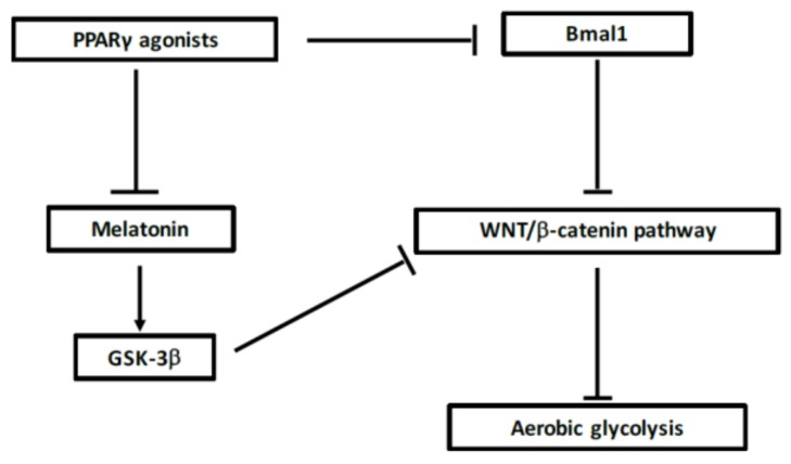
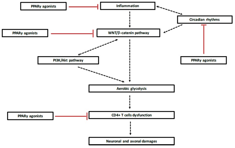
Demyelination in multiple sclerosis (MS) cells is the site of several energy metabolic abnormalities driven by dysregulation between the opposed interplay of peroxisome proliferator-activated receptor γ (PPARγ) and WNT/β-catenin pathways. We focus our review on the opposing interactions observed in demyelinating processes in MS between the canonical WNT/β-catenin pathway and PPARγ and their reprogramming energy metabolism implications. Demyelination in MS is associated with chronic inflammation, which is itself associated with the release of cytokines by CD4⁺ Th17 cells, and downregulation of PPARγ expression leading to the upregulation of the WNT/β-catenin pathway. Upregulation of WNT/β-catenin signaling induces activation of glycolytic enzymes that modify their energy metabolic behavior. Then, in MS cells, a large portion of cytosolic pyruvate is converted into lactate. This phenomenon is called the Warburg effect, despite the availability of oxygen. The Warburg effect is the shift of an energy transfer production from mitochondrial oxidative phosphorylation to aerobic glycolysis. Lactate production is correlated with increased WNT/β-catenin signaling and demyelinating processes by inducing dysfunction of CD4⁺ T cells leading to axonal and neuronal damage. In MS, downregulation of PPARγ decreases insulin sensitivity and increases neuroinflammation. PPARγ agonists inhibit Th17 differentiation in CD4⁺ T cells and then diminish release of cytokines. In MS, abnormalities in the regulation of circadian rhythms stimulate the WNT pathway to initiate the demyelination process. Moreover, PPARγ contributes to the regulation of some key circadian genes. Thus, PPARγ agonists interfere with reprogramming energy metabolism by directly inhibiting the WNT/β-catenin pathway and circadian rhythms and could appear as promising treatments in MS due to these interactions.

Keywords: PPARγ; WNT/β-catenin pathway; Warburg effect; aerobic glycolysis; circadian rhythms; clock genes; demyelination; energy metabolism; multiple sclerosis.


Conflict of interest statement

The authors declare that they have no competing interests.

Figures

Figure 1
Figure 1
The canonical WNT/β-catenin pathway. (A) Under physiological circumstances, the WNT “off state”, the cytosolic β-catenin is bound to its destruction complex, consisting of adenomatous polyposis coli (APC), AXIN and glycogen synthase kinase-3β (GSK-3β). After CK-1 phosphorylates on Ser45 residue, β-catenin is further phosphorylated on Thr41, Ser37, and Ser33 residues by GSK-3β. Then, phosphorylated β-catenin is degraded into the proteasome. Therefore, the cytosolic level of β-catenin is kept low in the absence of WNT ligands. If β-catenin is not present in the nucleus, the T-cell factor/lymphoid enhancer factor (TCF/LEF) complex cannot activate the target genes. Dickkopf (DKK) can inhibit the WNT/β-catenin pathway by binding to WNT ligands or low-density lipoprotein receptor-related protein 5/6 (LRP 5/6). (B) When WNT ligands bind to both Frizzled (FZD) and LRP 5/6, the WNT “on state”, Disheveled (DSH) is recruited and phosphorylated by FZD. Phosphorylated DSH in turn recruits AXIN, which dissociates the β-catenin destruction complex. Therefore, β-catenin escapes from phosphorylation and subsequently accumulates in the cytosol. The accumulated cytosolic β-catenin goes into the nucleus, where it binds to TCF/LEF and activates the transcription of target genes.
Figure 2
Figure 2
Aerobic glycolysis stimulation by activated canonical WNT/β-catenin pathway. Activation of the receptor tyrosine kinase (RTK) is required to take up enough glucose to cell survival. PI3K/Akt pathway is stimulated to maintain a sufficient ATP production through the metabolism of glucose. Glucose is transformed into pyruvate into the mitochondria for the oxidative phosphorylation process. However, during WNT activation, WNT signal transduction results in activation of c-Myc, lactate dehydrogenase A (LDH-A), pyruvate dehydrogenase kinase (PDK) and monocarboxylate transporter 1 (MCT-1). The WNT target genes cooperate to divert glycolytically derived pyruvate into lactate which is expelled out the cell by MCT-1. Moreover, c-Myc induces glutamine uptake and glutaminolysis to support mitochondrial integrity and aspartate production. Accumulation of cytosolic lactate involves several pathways such as nucleotide synthesis, lipid synthesis and cell division.
Figure 3
Figure 3
Circadian clock genes process. The clock is considered as a stimulatory loop, with the Bmal1/Clock heterodimer activating the transcription of Period (Per) and Cryptochrome (Cry) genes, and then a negative feedback loop with the Per/Cry heterodimer which translocates to the nucleus and then represses the transcription of the Clock and Bmal1 genes. An additional loop implicates the RORs and Rev Erbs factors with a positive feedback by retinoid-related orphan receptors (ROR) and a negative feedback by Rev Erbs. Arrows: activation; T bar: inhibition.
Figure 4
Figure 4
Schematic interaction between WNT, peroxisome proliferator-activated receptor γ (PPARγ) and circadian rhythms. PPARγ agonists can decrease Bmal1 expression. The knockout of Bmal1 leads to decrease WNT/β-catenin pathway activity and then in absence of initiation of aerobic glycolysis. In parallel, PPARγ agonists can decrease melatonin levels leading to activate GSK-3β activity, the main inhibitor of WNT pathway.
Figure 5
Figure 5
Potential PPARγ agonists treatment approach in demyelination. During acute phase, inflammation processes, activated by disruption of circadian rhythms, lead to release of several cytokines and pro-inflammatory factors which stimulate the canonical WNT/β-catenin pathway. Activation of the WNT ligands involves WNT target genes that are responsible for the initiation of the shunt of the tricarboxylic acid (TCA) resulting in aerobic glycolysis instead of oxidative phosphorylation. Lactate production, the main factor of energy metabolism alteration, and its release out the cells enhance CD4+ T cells dysfunction which aggravates MS pathogenesis, neuronal and axonal damages. Using PPARγ agonists could be interesting because of their four interactions in the demyelination cascade. First, PPARγ agonists directly inhibits neuroinflammation by inhibiting cytokines and inflammatory factors release. Secondly, PPARγ agonists can regulate circadian clocks, such as Bmal1, to decrease inflammatory factors release and to target WNT ligands. Third, their opposed interaction with the canonical WNT/β-catenin pathway can prevent the initiation of aerobic glycolysis process and then the energy metabolism reprogramming enable MS. At last, PPARγ agonists have neuroprotective effects by targeting CD4+ T cells to prevent neuronal and axonal damages. Arrow: activation; T bar: inhibition.

References

    1. Goverman J. Autoimmune T cell responses in the central nervous system. Nat. Rev. Immunol. 2009;9:393–407. doi: 10.1038/nri2550. - DOI - PMC - PubMed
    1. Trapp B.D., Nave K.-A. Multiple sclerosis: An immune or neurodegenerative disorder? Annu. Rev. Neurosci. 2008;31:247–269. doi: 10.1146/annurev.neuro.30.051606.094313. - DOI - PubMed
    1. Rasmussen S., Wang Y., Kivisäkk P., Bronson R.T., Meyer M., Imitola J., Khoury S.J. Persistent activation of microglia is associated with neuronal dysfunction of callosal projecting pathways and multiple sclerosis-like lesions in relapsing—Remitting experimental autoimmune encephalomyelitis. Brain J. Neurol. 2007;130:2816–2829. doi: 10.1093/brain/awm219. - DOI - PubMed
    1. Jadidi-Niaragh F., Mirshafiey A. Th17 cell, the new player of neuroinflammatory process in multiple sclerosis. Scand. J. Immunol. 2011;74:1–13. doi: 10.1111/j.1365-3083.2011.02536.x. - DOI - PubMed
    1. Didonna A., Oksenberg J.R. Genetic determinants of risk and progression in multiple sclerosis. Clin. Chim. Acta Int. J. Clin. Chem. 2015;449:16–22. doi: 10.1016/j.cca.2015.01.034. - DOI - PMC - PubMed