. 2018 Apr 16;14(4):e1007351.
doi: 10.1371/journal.pgen.1007351. eCollection 2018 Apr.

Cytokinin stabilizes WUSCHEL by acting on the protein domains required for nuclear enrichment and transcription

Affiliations

Cytokinin stabilizes WUSCHEL by acting on the protein domains required for nuclear enrichment and transcription

Stephen A Snipes et al. PLoS Genet. .

Abstract

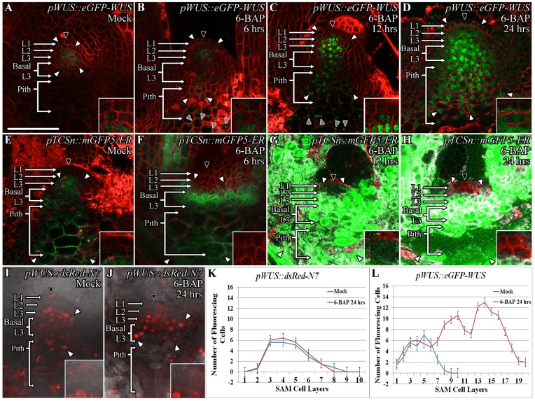
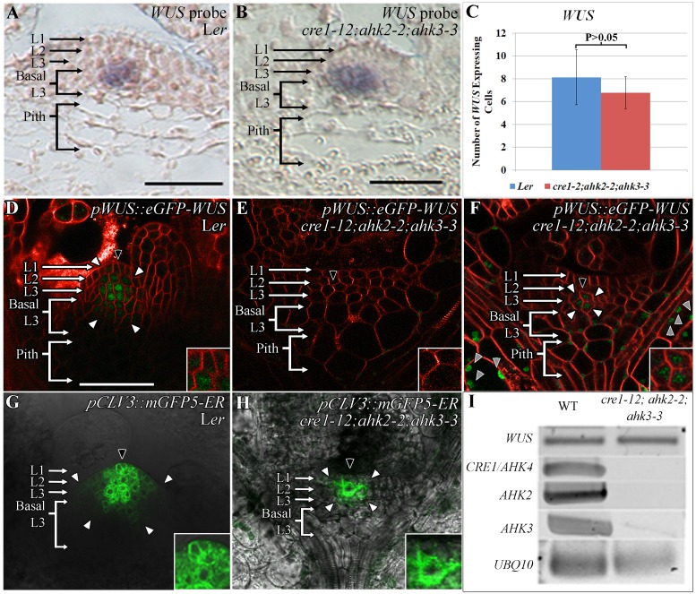
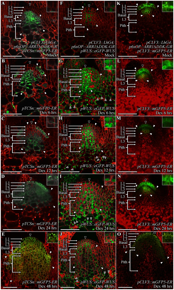
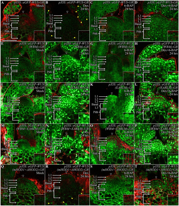
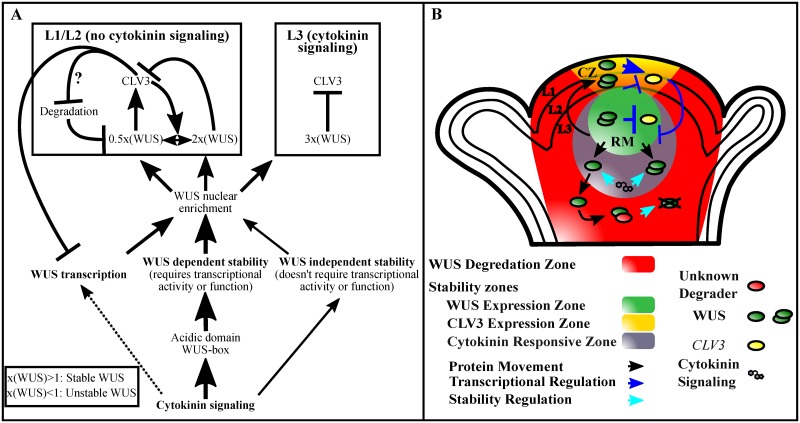
Concentration-dependent transcriptional regulation and the spatial regulation of transcription factor levels are poorly studied in plant development. WUSCHEL, a stem cell-promoting homeodomain transcription factor, accumulates at a higher level in the rib meristem than in the overlying central zone, which harbors stem cells in the shoot apical meristems of Arabidopsis thaliana. The differential accumulation of WUSCHEL in adjacent cells is critical for the spatial regulation and levels of CLAVATA3, a negative regulator of WUSCHEL transcription. Earlier studies have revealed that DNA-dependent dimerization, subcellular partitioning and protein destabilization control WUSCHEL protein levels and spatial accumulation. Moreover, the destabilization of WUSCHEL may also depend on the protein concentration. However, the roles of extrinsic spatial cues in maintaining differential accumulation of WUS are not understood. Through transient manipulation of hormone levels, hormone response patterns and analysis of the receptor mutants, we show that cytokinin signaling in the rib meristem acts through the transcriptional regulatory domains, the acidic domain and the WUSCHEL-box, to stabilize the WUS protein. Furthermore, we show that the same WUSCHEL-box functions as a degron sequence in cytokinin deficient regions in the central zone, leading to the destabilization of WUSCHEL. The coupled functions of the WUSCHEL-box in nuclear retention as described earlier, together with cytokinin sensing, reinforce higher nuclear accumulation of WUSCHEL in the rib meristem. In contrast a sub-threshold level may expose the WUSCHEL-box to destabilizing signals in the central zone. Thus, the cytokinin signaling acts as an asymmetric spatial cue in stabilizing the WUSCHEL protein to lead to its differential accumulation in neighboring cells, which is critical for concentration-dependent spatial regulation of CLAVATA3 transcription and meristem maintenance. Furthermore, our work shows that cytokinin response is regulated independently of the WUSCHEL function which may provide robustness to the regulation of WUSCHEL concentration.


Conflict of interest statement

The authors have declared that no competing interests exist.

Figures

Fig 1
Fig 1. Exogenous application of cytokinin leads to higher WUS protein accumulation which corresponds with the spatial pattern of induction of cytokinin response.
Side views of SAMs showing WUS protein (pWUS::eGFP-WUS) accumulation in 9 day old Mock-treated (A) and 6-BAP treated Ler plants for 6 hrs (B), 12 hrs (C), and 24 hrs (D). pTCSn::mGFP5-ER reporter expression in 9 day old Mock-treated (E) and 6-BAP treated Ler plants for 6 hrs (F), 12 hrs (G), and 24 hrs (H). The SAMs showing the expression of pWUS::dsRed-N7 transcriptional reporter which accumulates mostly in the apical L3 and Basal L3 layers upon Mock (I) and 6-BAP treatment for 24 hrs (J). A slight lateral expansion of WUS expression domain by 1–2 cell layers has been observed upon 6-BAP treatment for 24 hrs (J). The number of WUS positive cells in different SAM layers as measured by the expression of transcriptional reporter (pWUS::dsRed-N7) upon Mock and 6-BAP treatment for 24 hrs (K) (n = 12) is compared to the number of cells that accumulate WUS protein in different SAM layers as measured by the translational reporter (pWUS::eGFP-WUS) upon Mock and 6-BAP treatment for 24 hrs (n = 7) (L). Error bars for (K-L) represent standard error. The cell layers in SAMs are marked; the L1 and the L2 are monolayers. The multilayer L3 has been divided into the apical L3 layer and the basal L3 layers. The pith is located beneath the basal L3 layers. Insets for each image show the areas identified by black arrowheads at 4x zoom and white arrowheads show the boundaries of the reporter accumulation. Autofluorescence is denoted by grey arrowheads and is characterized by multiple foci in a single cell. eGFP and mGFP-ER (green) are overlaid on FM4-64 (red) plasma membrane stain in (A-H). The dsRed-N7 (red) is overlaid on DIC images (I-J). The scale bars = 50 μm for all images.
Fig 2
Fig 2. Lower WUS protein accumulation and altered CLV3 expression in cytokinin receptor mutants.
RNA in situ showing localization of WUS transcripts in Ler (A) and in cre1-12;ahk2-2;ahk3-3 (B) lines. The number of WUS expressing cells quantified from RNA in situ images in Ler and cre1-12;ahk2-2;ahk3-3. P>0.05 as determined by a Student’s t-test (C). WUS protein (pWUS::eGFP-WUS) accumulation in Ler (D) and in cre1-12;ahk2-2;ahk3-3 (E-F). cre1-12;ahk2-2;ahk3-3 plants showing no detectable pWUS::eGFP-WUS accumulation (E), a few pWUS::eGFP-WUS accumulating cells detected when imaged at 1.5x detector gain (F). The pCLV3::mGFP5-ER expression in Ler (G) and cre1-12;ahk2-2;ahk3-3 (H). Semi-quantitative RT-PCR of WUS and CRE1/AHK4, AHK2, and AHK3 transcripts in wild type and cre1-12;ahk2-2;ahk3-3 mutants (I). The cell layers in SAMs are marked; the L1 and the L2 are monolayers. The multilayer L3 has been divided into the apical L3 layer and the basal L3 layers. The pith is located beneath the basal L3 layers. Insets for each image show the areas identified by black arrowheads at 4x zoom and white arrowheads show the boundaries of reporter accumulation. Autofluorescence is denoted by grey arrowheads and is characterized by multiple foci in a single cell. eGFP (green) is overlaid on FM4-64 (red) plasma membrane stain in (D-F). mGFP5-ER is overlaid on DIC in (G-H). The scale bars = 50 μm for all images.
Fig 3
Fig 3. The ectopic activation of cytokinin signaling leads to higher WUS protein accumulation in SAMs, including the outer cell layers.
The SAM images of nine day old pCLV3::LhG4; p6xOP::ARR1ΔDDK-GR seedling showing pTCSn::mGFP5-ER reporter expression (A-E), WUS protein (pWUS::eGFP-WUS) accumulation (F-J), and CLV3 expression (pCLV3::mGFP5-ER) (K-O) in Mock-treated plants (A, F, K) and at 6 hrs (B, G, L), 12 hrs (C, H, M), 24 hrs (D, I, N), and 48hrs (E, J, O) after Dex treatment. The cell layers in SAMs are marked; the L1 and the L2 are monolayers. The multilayer L3 has been divided into the apical L3 layer and the basal L3 layers. The pith is located beneath the basal L3 layers. Insets for each image show the areas identified by black arrowheads at 4x zoom and white arrowheads show the boundaries of reporter accumulation. Autofluorescence is denoted by grey arrowheads and is characterized by multiple foci in a single cell. mGFP-ER and eGFP (green) are overlaid on FM4-64 (red) plasma membrane stain. The scale bars = 50 μm for all images.
Fig 4
Fig 4. Cytokinin can offset ectopic WUS overexpression induced instability, which depends on the degron-like function of the WUS-box but does not require transcriptional activity of WUS.
The SAMs showing the accumulation of ubiquitously expressed wild type (p35S::eGFP-WUS-GR), WUS-box mutant (p35S::eGFP-WUS (WBM)-GR), EAR-like domain mutant (p35S::eGFP-WUS (EARLM)-GR), double mutants of WBM mutant with the EARLM domain mutant (p35S::eGFP-WUS (WBM+EARLM)-GR), and double mutants of mHOD1 and ΔHOD2 (p35::eGFP-WUS (mHOD1+ΔHOD2)-GR) versions in Mock-treated (A, E, I, M, Q), Dex-treated for 24 hrs (B, F, J, N, R), 6-BAP-treated for 24 hrs (C, G, K, O, S), and combined Dex+6-BAP treated for 24 hrs (D, H, L, P, T) plants. 6-BAP treatment alone led to increase in cytoplasmically-localized WUS in wild type (C), EARLM (K), and the double mutants of mHOD1 and ΔHOD2 (S) while the WBM (G) and the double mutants of WBM and EARLM (O) did not reveal a striking increase in protein levels. Dex-treatment alone led to a dramatically lower levels of protein accumulation in cells located in the SAM (shown by the black arrowhead) in comparison to cells located in the developing leaves and the pith (yellow arrowheads) in wild type (B) and the double mutants of mHOD1 and ΔHOD2 (R) while the combined Dex and 6-BAP treatment improved protein accumulation both in wild type (D) and the double mutants of mHOD1 and ΔHOD2 (T). The Dex treatment of the WBM (F), EARLM (J) and the double mutants of WBM and EARLM (N) led to stable nuclear accumulation, though the WBM (F) did not show tight nuclear localization seen with the EARLM (J) and the WBM plus EARLM double mutants (N). The combined Dex and 6-BAP treatment (H, L, P) did not reveal a striking difference from that of the Dex alone treatment (F, J, N) in all three WUS forms. The cell layers in SAMs are marked; the L1 and the L2 are monolayers. The multilayer L3 has been divided into the apical L3 layer and the basal L3 layers. The pith is located beneath the basal L3 layers. Insets for each image show the areas identified by black arrowheads at 4x zoom. eGFP (green) is overlaid on FM4-64 (red) plasma membrane stain. The scale bars = 50 μm for all images.
Fig 5
Fig 5. Cytokinin signaling acts on the transcriptional regulatory domains to stabilize WUS.
Accumulation of various mutant forms of the WUS protein, expressed from the WUS promoter, 24 hrs after Mock (A-F) and 6-BAP treatment (G-L). pWUS::eGFP-WUS (A, G), pWUS::eGFP-WUS (229–292) (B, H), pWUS::eGFP-WUS (ADM) (C, I), pWUS::eGFP-WUS (WBM) (D, J), pWUS::eGFP-WUS (EARLM) (E, K) and pWUS::eGFP-WUS (WBM+EARLM) (F, L). A sketch showing the functions of WUS domains and associated amino acid changes in the mutant versions (M). Quantification of the relative fluorescence levels (N-S) and the number of fluorescing cells (T-Y) in Mock and 6-BAP treated plants. Error bars for (N-Y) represent standard error. The cell layers in SAMs are marked; the L1 and the L2 are monolayers. The multilayer L3 has been divided into the apical L3 layer and the basal L3 layers. The pith is located beneath the basal L3 layers. Insets for each image show the areas identified by black arrowheads at 4x zoom, white arrowheads show the boundaries of reporter accumulation, and yellow arrowheads point to WUS protein accumulation in developing leaves. Autofluorescence is denoted by grey arrowheads and is characterized by multiple foci in a single cell. eGFP (green) is overlaid on FM4-64 (red) plasma membrane stain. The scale bars = 50 μm for all images.
Fig 6
Fig 6. Transcriptional activity/functional WUS is required for stabilizing the WUS protein.
The pWUS::eGFP-WUS (mHOD1+ΔHOD2) accumulation in wild type SAMs upon Mock (A), 6 hrs (B), and 24 hrs (C) of 6-BAP treatments. Note the diffuse accumulation of WUS and the response to 6-BAP treatments, particularly in the pith cells in (B-C). The pWUS::eGFP-WUS (mHOD1+ΔHOD2) accumulation in wus-1 SAMs upon Mock (D), 6 hrs (E) and 24 hrs (F) of 6-BAP treatments. Note the poor accumulation of WUS in (D) and the much weaker response to 6-BAP treatments in (E-F). Cytokinin signaling response from the pTCS::mGFP5-ER in wus-1 SAMs after Mock (G), 6 hrs (H), and 24 hrs (I) of 6-BAP treatments. The cytokinin response is clearly evident even after 6 hrs of treatment compared to Mock, demonstrating that cytokinin signaling is normal in wus-1 SAMs and that the transcriptional activity/function of WUS is required for WUS stabilization. (A-F) are expressed from pWUS. The cell layers in SAMs are marked; the L1 and the L2 are monolayers. The multilayer L3 has been divided into the apical L3 layer and the basal L3 layers. The pith is located beneath the basal L3 layers. Insets for each image show the areas identified by black arrowheads at 4x zoom and white arrowheads show boundaries of the reporter accumulation. Autofluorescence is denoted by grey arrowheads and is characterized by multiple foci in a single cell. eGFP (green) is overlaid on FM4-64 (red) plasma membrane stain. The scale bar is 50 μm for all images.
Fig 7
Fig 7. Summary of cytokinin effects on WUS and a model for the role of cytokinin in WUS-mediated regulation of the stem cell niche.
(A) Cytokinin at higher level promotes WUS transcription. However, within the physiological range it stabilizes WUS protein by acting on the acidic domain and the WUS-box. The WUS-box is also required for nuclear retention and transcriptional regulation. A small amount of this stabilization can occur through a WUS-independent mechanism, while the majority of the cytokinin-mediated WUS stabilization requires WUS function. The consequent nuclear enrichment of WUS (3X) in the RM may mask degron activity of the WUS-box, leading to stability which in turn represses CLV3 transcription. The L1 and the L2 layers of central part of the SAM are devoid of cytokinin signaling, where the degron activity of the WUS-box results in unstable WUS accumulation. However, CLV3 signaling partially offsets this instability in the L1 and the L2 layers. A relatively higher WUS (2X) represses CLV3 which destabilizes WUS (0.5X), which in turn activates CLV3, thus forming a feedback loop at the post-translational level. The range of WUS levels represented (0.5X to 3X) are only relative. (B) A schematic showing the spatial representation of interactions that lead to stable and unstable WUS accumulation in space. Degradation system (red) is ubiquitously present. The cytokinin responsive zone (grey) in the RM stabilizes WUS protein. The wild type CLV3 signaling strength stabilizes WUS in the L1 and the L2 layers, while a lower CLV3 destabilizes WUS. Thus oscillating WUS levels mediate concentration-dependent activation and repression of CLV3 transcription forming a self-sustaining feedback loop. WUS (green) and CLV3 (Yellow) expression zones are marked.

References

    1. Rushlow CA, Han K, Manley JL, Levine M. The graded distribution of the dorsal morphogen is initiated by selective nuclear transport in Drosophila. Cell. 1989;59: 1165–1177. - PubMed
    1. Nellen D, Burke R, Struhl G, Basler K. Direct and long-range action of a DPP morphogen gradient. Cell. 1996;85: 357–368. - PubMed
    1. Briscoe J, Ericson J. The specification of neuronal identity by graded Sonic Hedgehog signalling. Semin Cell Dev Biol. 1999;10: 353–362. doi: 10.1006/scdb.1999.0295 - DOI - PubMed
    1. Steeves TA, Sussex IM. Patterns in plant development. Cambridge University Press; 1989.
    1. Reddy GV, Heisler MG, Ehrhardt DW, Meyerowitz EM. Real-time lineage analysis reveals oriented cell divisions associated with morphogenesis at the shoot apex of Arabidopsis thaliana. Development. 2004;131: 4225–4237. doi: 10.1242/dev.01261 - DOI - PubMed

Publication types

MeSH terms

LinkOut - more resources