. 2018 Jun 1;103(6):2126-2135.
doi: 10.1210/jc.2017-01562.

Increased Chromogranin A-Positive Hormone-Negative Cells in Chronic Pancreatitis

Affiliations

Increased Chromogranin A-Positive Hormone-Negative Cells in Chronic Pancreatitis

Abu Saleh Md Moin et al. J Clin Endocrinol Metab. .

Abstract

Context: Chronic pancreatitis (CP) is characterized by inflammation, fibrosis, and a loss of pancreatic acinar cells, which can result in exocrine and eventually endocrine deficiency. Pancreatitis has been reported to induce formation of new endocrine cells (neogenesis) in mice. Our recent data have implicated chromogranin A-positive hormone-negative (CPHN) cells as potential evidence of neogenesis in humans.

Objective: We sought to establish if CPHN cells were more abundant in CP in humans.

Design, setting, and participants: We investigated the frequency and distribution of CPHN cells and the expression of the chemokine C-X-C motif ligand 10 (CXCL10) and its receptor chemokine C-X-C motif receptor 3 in pancreas of nondiabetic subjects with CP.

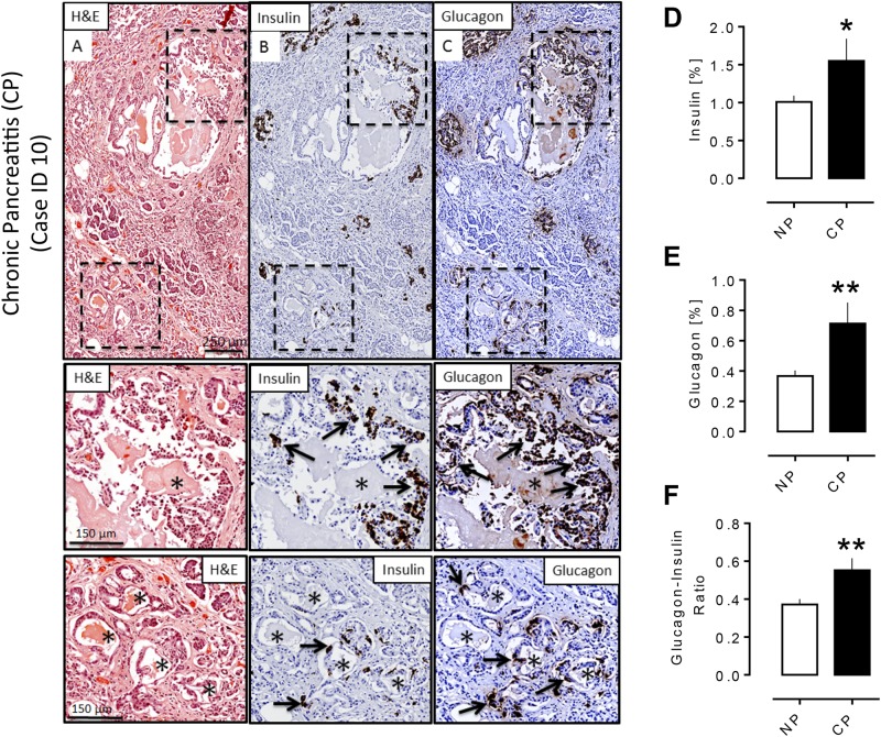
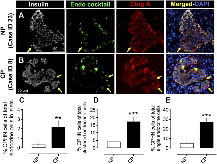
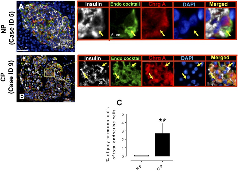
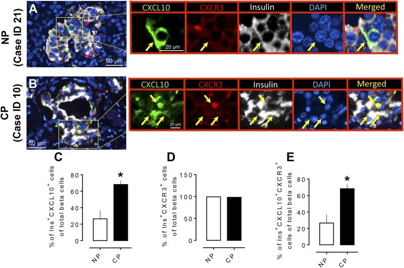
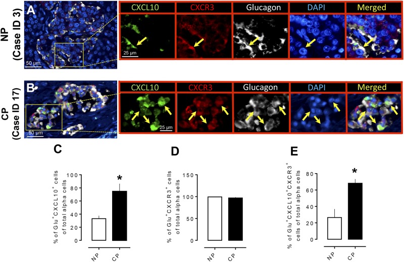
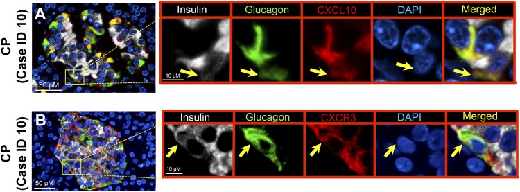
Results: CPHN cell frequency in islets was increased sevenfold in CP [2.1% ± 0.67% vs 0.35% ± 0.09% CPHN cells in islets, CP vs nonpancreatitis (NP), P < 0.01], as were the CPHN cells found as scattered cells in the exocrine areas (17.4 ± 2.9 vs 4.2 ± 0.6, CP vs NP, P < 0.001). Polyhormonal endocrine cells were also increased in CP (2.7 ± 1.2 vs 0.1 ± 0.04, CP vs NP, % of polyhormonal cells of total endocrine cells, P < 0.01), as was expression of CXCL10 in α and β cells.

Conclusion: There is increased islet endogenous expression of the inflammation marker CXCL10 in islets in the setting of nondiabetic CP and an increase in polyhormonal (insulin-glucagon expressing) cells. The increase in CPHN cells in CP, often in a lobular distribution, may indicate foci of attempted endocrine cell regeneration.


Figures

Figure 1.
Figure 1.
Pathology of pancreas in a subject with CP. Adjacent sections of pancreas from a subject with CP (40-year-old male, BMI 34.6) stained for (A) hematoxylin and eosin (H&E), (B) insulin (diaminobenzidine) with hematoxylin counterstain, and (C) glucagon (diaminobenzidine) with hematoxylin counterstain, showing typical features of CP (i.e., fibrosis, loss of acinar tissue, dilated pancreatic ducts, and areas of acinar-to ductal metaplasia). Insets show an increase of insulin- and glucagon-positive cells (arrows) in ductal epithelium (upper) and in acinar-to-ductal metaplasia (lower) with a preponderance of glucagon-positive cells in both locations. (D, E) Percentage of (D) insulin and (E) glucagon fractional area in the subjects with CP and without CP (NP). Both insulin and glucagon fractional area were increased in the CP group (insulin area %: 1.55 ± 0.29 vs 1.01 ± 0.08, CP vs NP, *P < 0.05; glucagon area %: 0.71 ± 0.14 vs 0.37 ± 0.04, CP vs NP, **P < 0.005). (F) The glucagon/insulin ratio was increased in subjects with CP (0.55 ± 0.06 vs 0.37 ± 0.03, CP vs NP, **P < 0.005). CP, n = 19; NP, n = 38. *Ductules in an area of acinar-to-ductal metaplasia. Scale bar, 250 μm in lower-power views, 150 μm in insets.
Figure 2.
Figure 2.
Prevalence of hormone-negative endocrine cells in subjects with CP. Examples of CPHN cells in pancreas from (A) an NP subject and (B) a subject with CP. Individual layers stained for insulin (white), endocrine cocktail (glucagon, somatostatin, pancreatic polypeptide, and ghrelin) (green), chromogranin A (red), and 4′,6-diamidino-2-phenylindole (DAPI) (blue) are shown along with the merged image. Arrows indicate CPHN cells. Scale bars, 50 μm for both (A) and (B). The frequency of nonhormone-expressing endocrine cells was increased in (C) islets in subjects with CP (2.2 ± 0.6 vs 0.34 ± 0.1 CPHN cells per islet cross-section, CP vs NP, **P < 0.0001), in (D) scattered clusters (17.4 ± 2.9 vs 4.2 ± 0.5 cells/mm2, CP vs NP, ***P < 0.0001), and (E) as single cells (27.4 ± 3.8 vs 5.2 ± 1.0 cells/mm2, CP vs NP, **P < 0.001). CP, n = 12; NP, n = 24.
Figure 3.
Figure 3.
Expression of polyhormonal cells in islets in human subjects with CP. Representative merged images showing coexpression of insulin, endocrine cocktail, and chromogranin A along with 4′,6-diamidino-2-phenylindole (DAPI) in the pancreas from (A) an NP subject and (B) a subject with CP. Insets: Higher magnification of the indicated region in the low-power images; arrows indicate the polyhormonal cells that express insulin (white), endocrine cocktail (glucagon, somatostatin, pancreatic polypeptide, and ghrelin) (green), chromogranin A (red), and DAPI (blue). Individual layers are shown along with the merged image. Scale bars, 50 μm for both (A) and (B) in lower-power views, 5 μm (A) and 25 μm (B) in insets. (C) The percentage of polyhormonal cells of total endocrine cells in CP was higher compared with NP. **P < 0.01. CP, n = 13; NP, n = 22.
Figure 4.
Figure 4.
Expression of the chemokine CXCL10 and its receptor (CXCR3) in pancreatic β cells in CP. Representative merged images showing coexpression of CXCL10 and CXCR3 with insulin in the pancreas from (A) an NP subject and (B) a subject with CP. Insets: Higher magnification of the indicated region in the low-power images, with arrows indicating the cells that express CXCL10 (green), CXCR3 (red), insulin (white), 4′,6-diamidino-2-phenylindole (DAPI) (blue), and merged (yellow). Scale bar, 50 μm in lower-power views, 20 μm in insets. (C) The percentage of β cells that express CXCL10 was higher in CP compared with NP. (D) There was no change in the frequency of β cells that express the receptor (CXCR3) in CP or NP. (E) The percentage of Ins+CXCL10+CXCR3+ (triple-positive cells) was higher in CP compared with NP. *P < 0.05. CP, n = 4; NP, n = 4.
Figure 5.
Figure 5.
Expression of the chemokine CXCL10 and its receptor (CXCR3) in pancreatic α cells in CP. Representative merged images showing coexpression of CXCL10 and CXCR3 with glucagon in the pancreas from (A) an NP subject and (B) a subject with CP. Insets: Higher magnification of the indicated region in the low-power images, with arrows indicating the cells that express CXCL10 (green), CXCR3 (red), glucagon (white), 4′,6-diamidino-2-phenylindole (DAPI) (blue), and merged (yellow). Scale bar, 50 μm in lower-power views, 25 μm in insets. (C) The percentage of α cells that express CXCL10 was higher in CP compared with NP. (D) There was no change of percentage of α cells that express receptor (CXCR3) in CP or in NP. (E) The percentage of Glu+CXCL10+CXCR3+ (triple-positive cells) was higher in CP compared with NP. *P < 0.05. CP, n = 4; NP, n = 4.
Figure 6.
Figure 6.
Expression of chemokine (CXCL10) and its receptor (CXCR3) in α-β double-positive cells. Representative merged images showing (A) coexpression of insulin-glucagon-CXCL10 and (B) insulin-glucagon-CXCR3 in subjects with CP. Insets: Higher magnification of the indicated region in the low-power images, with arrows indicating (A) the cells that express insulin (white), glucagon (green), CXCL10 (red), 4′,6-diamidino-2-phenylindole (DAPI) (blue), and merged (yellow) and (B) insulin (white), glucagon (green), CXCR3 (red), DAPI (blue), and merged (yellow). Scale bar, 50 μm in lower-power views, 10 μm in insets.

References

    1. Klöppel G, Maillet B. The morphological basis for the evolution of acute pancreatitis into chronic pancreatitis. Virchows Arch A Pathol Anat Histopathol. 1992;420(1):1–4. - PubMed
    1. Steer ML, Waxman I, Freedman S. Chronic pancreatitis. N Engl J Med. 1995;332(22):1482–1490. - PubMed
    1. Sarner M, Cotton PB. Classification of pancreatitis. Gut. 1984;25(7):756–759. - PMC - PubMed
    1. Lankisch PG, Löhr-Happe A, Otto J, Creutzfeldt W. Natural course in chronic pancreatitis: pain, exocrine and endocrine pancreatic insufficiency and prognosis of the disease. Digestion. 1993;54(3):148–155. - PubMed
    1. Ammann RW, Akovbiantz A, Largiader F, Schueler G. Course and outcome of chronic pancreatitis. Longitudinal study of a mixed medical-surgical series of 245 patients. Gastroenterology. 1984;86(5 Pt 1):820–828. - PubMed

Publication types