. 2018 Dec 1;10(6):503-514.
doi: 10.1093/jmcb/mjy024.

EMC10 governs male fertility via maintaining sperm ion balance

Affiliations

EMC10 governs male fertility via maintaining sperm ion balance

Yuchuan Zhou et al. J Mol Cell Biol. .

Abstract

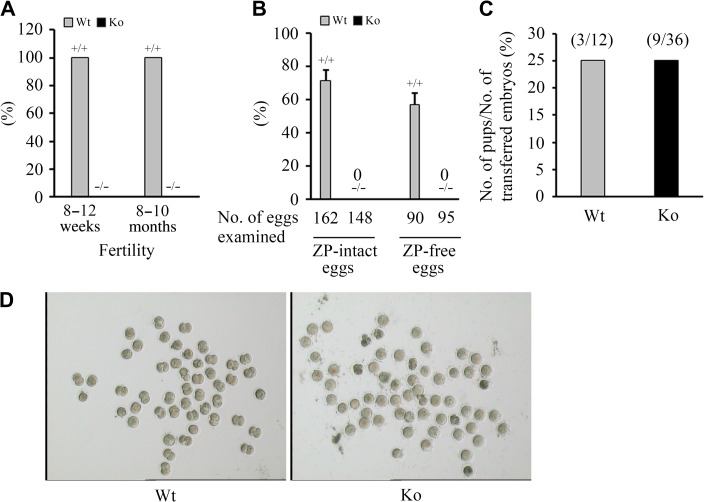
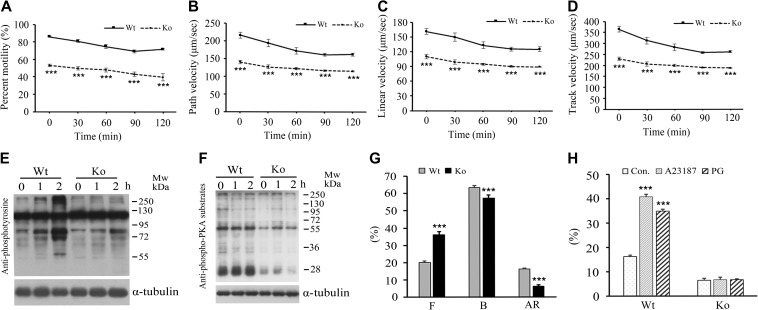
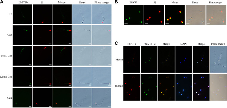
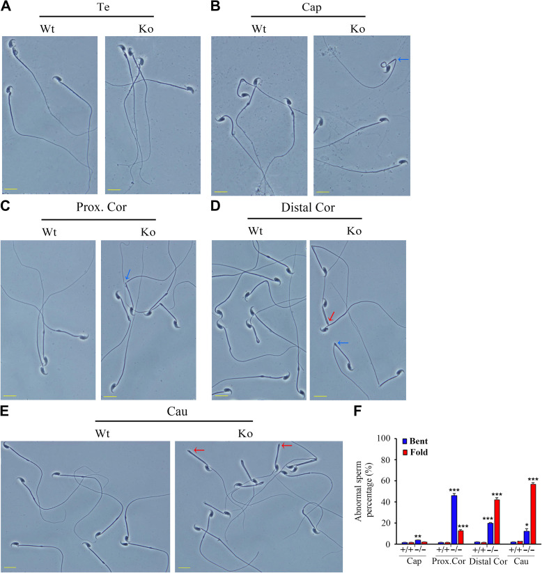
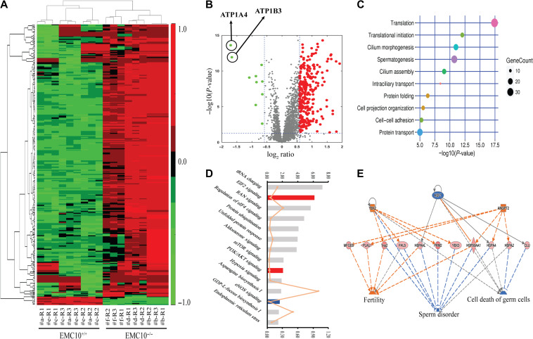
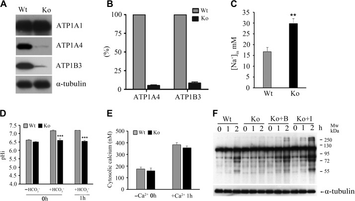
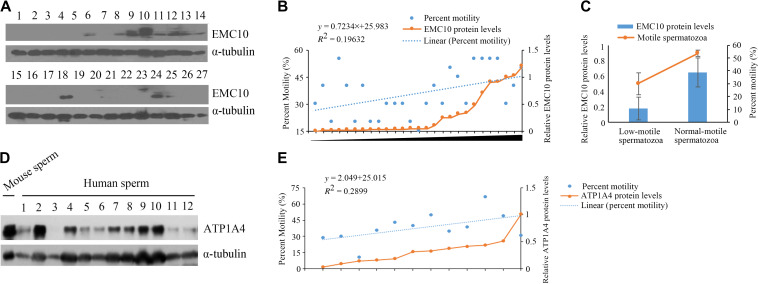
Infertility is a severe public health problem worldwide that prevails up to 15% in reproductive-age couples, and male infertility accounts for half of total infertility. Studies on genetically modified animal models have identified lots of genes involved in the pathogenesis of male infertility. The underlying causes, however, remain largely unclear. In this study, we provide evidence that EMC10, one subunit of endoplasmic reticulum (ER) membrane protein complex (EMC), is required for male fertility. EMC10 is significantly decreased in spermatozoa from patients with asthenozoospermia and positively associated with human sperm motility. Male mice lacking Emc10 gene are completely sterile. Emc10-null spermatozoa exhibit multiple defects including abnormal morphology, decreased motility, impaired capacitation, and impotency of acrosome reaction, thereby which are incapable of fertilizing intact or ZP-free oocytes. However, intracytoplasmic sperm injection could rescue this defect caused by EMC10 deletion. Mechanistically, EMC10 deficiency leads to inactivation of Na/K-ATPase, in turn giving rise to an increased level of intracellular Na+ in spermatozoa, which contributes to decreased sperm motility and abnormal morphology. Other mechanistic investigations demonstrate that the absence of EMC10 results in a reduction of HCO3- entry and subsequent decreases of both cAMP-dependent protein kinase A substrate phosphorylation and protein tyrosine phosphorylation. These data demonstrate that EMC10 is indispensable to male fertility via maintaining sperm ion balance of Na+ and HCO3-, and also suggest that EMC10 is a promising biomarker for male fertility and a potential pharmaceutical target to treat male infertility.


Figures

Figure 1
Figure 1
Effect of Emc10 disruption on male fertility. (A) Fertility of Emc10+/+ (Wt) and Emc10−/− (Ko) male mice (n = 18). (B) Analysis of in vitro fertilization of spermatozoa from Wt and Ko mice with intact and ZP-free oocytes, respectively (n = 3). Data are presented as mean ± SEM. (C) Analysis of the fertilizing ability after ICSI with spermatozoa from Wt and Ko mice. (D) Images of in vitro fertilization showing oocytes and two-cell embryos.
Figure 2
Figure 2
Effect of Emc10 disruption on sperm function. (A) The percentage of motile spermatozoa from wild-type (Wt) and Emc10-null (Ko) mice during 2-h incubation in medium (n = 4). (BD) Comparison of path velocity (B), linear velocity (C), and track velocity (D) of spermatozoa from Wt and Ko mice (n = 4). (E and F) Measurement of capacitation-associated protein tyrosine phosphorylation (E) and PKA substrate phosphorylation (F) of spermatozoa from Wt and Ko mice. Spermatozoa were incubated in capacitating medium and collected for western blot per hour. α-tubulin served as the loading control. The western blot is representative of four independent experiments. (G) The percentage of uncapacitated (F pattern), capacitated (B pattern), and acrosome-reacted (AR pattern) spermatozoa from Wt and Ko mice. (H) Examination of the acrosome reaction induced by A23187 or progesterone in sperm from Wt and Ko mice (n = 7). Data are presented as mean ± SEM. ***P < 0.001 when compared with respective controls.
Figure 3
Figure 3
The spatial distribution of EMC10 in mouse and human spermatozoon. (A) Immunostaining of EMC10 protein (green) on mouse spermatozoa from testis and different regions of the epididymis. Te, testis; Cap, caput epididymidis; Prox.Cor, proximal corpus epididymidis; Distal.Cor, distal corpus epididymidis; Cau, cauda epididymidis. (B) Immunostaining of EMC10 (green) on human spermatozoa. Nuclei were labeled with PI (red). (C) Immunofluorescent analysis of the localization of EMC10 protein (red) and the acrosome marker PNA-FITC (green) demonstrating the cellular localization of EMC10 in the acrosomal domain of spermatozoa from wild-type mice and humans. Nuclei were labeled with DAPI (blue). Scale bar, 10 μm.
Figure 4
Figure 4
Morphological defects of Emc10−/− spermatozoa. (AE) Phase contrast images of spermatozoa collected from the testis (A), caput (B), proximal corpus (C), distal corpus epididymidis (D), and cauda epididymidis (E) from wild-type (Wt) and Emc10-null (Ko) mice. Scale bar, 10 μm. (F) Quantification analysis of sperm with abnormal structure in different parts of the epididymis from Wt and Ko mice (n = 3). Data are presented as mean ± SEM, *P < 0.05, **P < 0.01, and ***P < 0.001 compared with respective controls. Te, testis; Cap, caput; Prox.Cor, proximal corpus; Distal.Cor, distal corpus; Cau, cauda epididymidis. Blue arrow, spermatozoa with bent tails; red arrow, spermatozoa with fold tails.
Figure 5
Figure 5
Proteomic analysis on the spermatozoa from wild-type and Emc10-null mice. (A) Heat map of 327 significantly increased or decreased proteins (>1.5-fold) in the spermatozoa of wild-type (Emc10+/+) and Emc10−/− mice. Red represents for the upregulated proteins and green for the downregulated proteins. Values were normalized to log2-fold difference. The false discovery rate is <0.10. (B) Volcano plot of 327 significantly altered proteins (>1.5-fold) in the spermatozoa of Emc10−/− mice compared with wild-type controls. Red, upregulation; green, downregulation. (C) GO term enrichment analysis. The 327 significantly changed proteins were analyzed to identify the GO biological processes from the DAVID website (http://david.abcc.ncifcrf.gov/). A R-Script diagram was generated to visualize the top 10 significantly altered biological processes in Emc10−/− spermatozoa. (D) Distribution of top 14 enriched pathways in the filtered data set of detected proteins according to the analysis by IPA (www.ingenuity.com). Fisher’s test was applied and proteins with >1.5-fold change in abundance were included. The top coordinates are –log10-transformed P-values. The bottom coordinates are the ratio of differentially expressed proteins and the number of proteins in the indicated pathway. Red, pathways were predicted to be significantly activated; blue, pathways predicted to be significantly downregulated; gray, pathways without predicted results. (E) The predicted work modules based on the 327 significantly altered proteins (>1.5-fold) in the sperm of Emc10−/− mice. Orange labeling indicates increased measurement (e.g. YBX2, ANGPT2) or the effect predicted to be activated (e.g. fertility), while blue characters represent decreased measurement (e.g. HSF1) or the effect predicted to be inhibited (e.g. sperm disorder, cell death of germ cells). The dashed lines indicate the predicted interactions between network proteins, and the directions of the interaction are indicated by orange arrows (activation), blue-blocked lines (inhibition), or gray lines (effect not predicted).
Figure 6
Figure 6
Effect of Emc10 deletion on ATPase expression and ionic equilibrium in spermatozoa. (A) Determination of ATP1A1, ATP1A4, and ATP1B3 protein levels in spermatozoa from wild-type (Wt) and Emc10-null (Ko) mice. α-tubulin was used as the protein loading control. The western blot shows a representative result of six independent experiments. (B) Quantitative analysis of ATP1A4 and ATP1B3 protein levels based on densitometry of western blot in A. (C) Intracellular Na+ concentration in spermatozoa from Wt and Ko mice. (D) Determination of intracellular pH in Wt and Ko spermatozoa incubated in HCO3-free medium or HCO3-containing medium for 0 or 1 h. (E) Determination of intracellular Ca2+ in Wt and Ko spermatozoa incubated in Ca2+-free or Ca2+-containing medium. (F) Detection of capacitation-associated protein tyrosine phosphorylation of Ko spermatozoa incubated in capacitating medium in the presence of 1 mM 8-Bromo-cAMP or 100 μM IBMX. A representative western blot of four independent experiments is shown. α-tubulin was used as the protein loading control. B, 8-Bromo-cAMP; I, IBMX. Data are presented as mean ± SEM, n = 4, **P < 0.01, ***P < 0.001, when compared with control.
Figure 7
Figure 7
The correlation of EMC10 or ATP1A4 protein level with sperm motility in humans. (A, B, D, and E) Immunoblot (IB) analysis was performed to determine the levels of EMC10 (A) and ATP1A4 (D) in sperm protein derived from asthenozoospermic patients and male controls. The scatter diagram shows the motile spermatozoa percentage and the relative protein levels of EMC10 (B) or ATP1A4 (E). The percentage of motile spermatozoa was quantified by ImageJ software (v2.1). The linear regression analysis was analyzed by using the Statistical Package for the Social Sciences (SPSS 20.0) software program. (C) The histogram shows motile sperm percentage and the relative protein levels of EMC10. Data are presented as mean ± SD, **P < 0.01.

References

    1. Abou-haila, A., and Tulsiani, D.R. (2009). Signal transduction pathways that regulate sperm capacitation and the acrosome reaction. Arch. Biochem. Biophys. 485, 72–81. - PubMed
    1. Baker, M.A., Hetherington, L., and Aitken, R.J. (2006). Identification of SRC as a key PKA-stimulated tyrosine kinase involved in the capacitation-associated hyperactivation of murine spermatozoa. J. Cell Sci. 119, 3182–3192. - PubMed
    1. Christianson, J.C., Olzmann, J.A., Shaler, T.A., et al. . (2011). Defining human ERAD networks through an integrative mapping strategy. Nat. Cell Biol. 14, 93–105. - PMC - PubMed
    1. Cooper, T.G., Yeung, C.H., Wagenfeld, A., et al. . (2004). Mouse models of infertility due to swollen spermatozoa. Mol. Cell. Endocrinol. 216, 55–63. - PubMed
    1. Darszon, A., Acevedo, J.J., Galindo, B.E., et al. . (2006). Sperm channel diversity and functional multiplicity. Reproduction 131, 977–988. - PubMed

Publication types