. 2018 Jun 2;500(2):423-428.
doi: 10.1016/j.bbrc.2018.04.093. Epub 2018 Apr 17.

Biophysical characterization of actin bundles generated by the Chlamydia trachomatis Tarp effector

Affiliations

Biophysical characterization of actin bundles generated by the Chlamydia trachomatis Tarp effector

Susmita Ghosh et al. Biochem Biophys Res Commun. .

Abstract

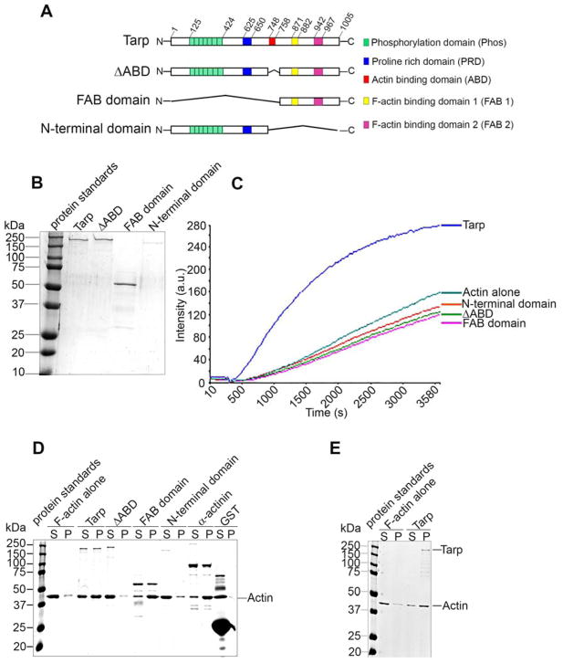
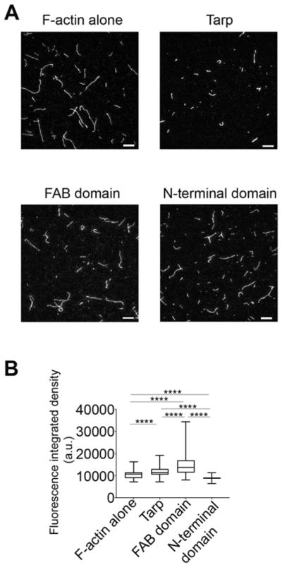
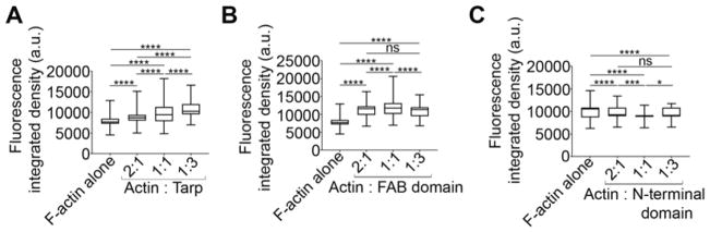
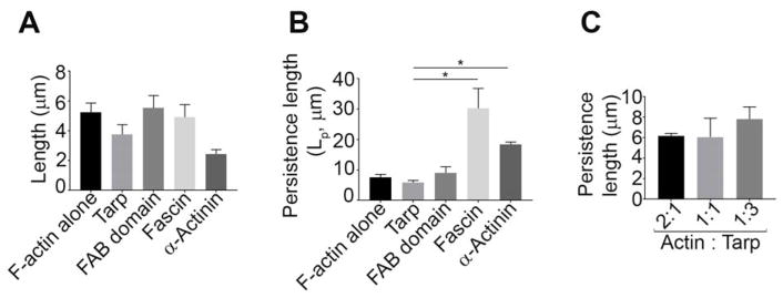
Chlamydia trachomatis entry into host cells is mediated by pathogen-directed remodeling of the actin cytoskeleton. The chlamydial type III secreted effector, translocated actin recruiting phosphoprotein (Tarp), has been implicated in the recruitment of actin to the site of internalization. Tarp harbors G-actin binding and proline rich domains required for Tarp-mediated actin nucleation as well as unique F-actin binding domains implicated in the formation of actin bundles. Little is known about the mechanical properties of actin bundles generated by Tarp or the mechanism by which Tarp mediates actin bundle formation. In order to characterize the actin bundles and elucidate the role of different Tarp domains in the bundling process, purified Tarp effectors and Tarp truncation mutants were analyzed using Total Internal Reflection Fluorescence (TIRF) microscopy. Our data indicate that Tarp mediated actin bundling is independent of actin nucleation and the F-actin binding domains are sufficient to bundle actin filaments. Additionally, Tarp-mediated actin bundles demonstrate distinct bending stiffness compared to those crosslinked by the well characterized actin bundling proteins fascin and alpha-actinin, suggesting Tarp may employ a novel actin bundling strategy. The capacity of the Tarp effector to generate novel actin bundles likely contributes to chlamydia's efficient mechanism of entry into human cells.

Keywords: Actin bundles; Bending persistence length; Chlamydia trachomatis; Cytoskeleton; Effector; Tarp.


Conflict of interest statement

Conflict of interest statement

The authors declare that the research was conducted in the absence of any commercial or financial relationships that could be construed as a potential conflict of interest. The content is solely the responsibility of the authors and does not necessarily represent the official views of the National Institutes of Health.

Figures

Figure 1
Figure 1
The Tarp FAB domain is sufficient to bundle actin. (A) Schematics of Tarp proteins utilized in this study indicating the locations of the tyrosine rich repeat phosphorylation domain (green boxes), the proline rich domain (blue box), G-actin binding domain (red box), and F-actin binding domains 1 (yellow box) and 2 (pink box). The numbers indicate amino acid positions encoded within the C. trachomatis tarP gene. The “∧” indicates amino acids deleted from the wild type sequence. Mutant Tarp clones included Tarp lacking the G-actin binding domain (ΔABD) as well as Tarp fragments representing only the F-actin binding domains (FAB domains) or a Tarp truncation excluding all known actin (both G- and F-) binding domains (N-terminal domain). (B) Purified Tarp and Tarp mutants depicted in (A). (C) Tarp proteins described in panels A and B were assessed in pyrene actin polymerization assays. Tarp proteins were incubated with monomeric pyrene-labeled actin. An increase in actin polymerization after the addition of polymerization buffer at 300 s was measured as arbitrary fluorescence intensity [Intensity (a.u.)] over time [Time (s)]. The data are representative of three repeated experiments. (D) Purified recombinant Tarp proteins were incubated with preformed filamentous actin (F-actin) and isolated by low-speed centrifugation. Protein supernatants (S) and pellets (P) were resolved by SDS-PAGE and visualized by Coomassie blue staining. α-actinin and GST served as positive and negative controls, respectively. (E) Globular actin (G-actin) along with polymerization buffer was incubated with (Tarp) or without (F-actin alone) purified recombinant Tarp and isolated by low-speed centrifugation. Protein supernatants (S) and pellets (P) were resolved by SDS-PAGE and visualized by Coomassie blue staining.
Figure 2
Figure 2
Tarp produces actin structures which have higher fluorescence intensities compared to actin filaments alone as determined by TIRF microscopy image analysis. (A) Representative TIRF images of rhodamine-labeled actin filaments or bundles assembled with Tarp or Tarp mutant proteins (Scale bar 10 μm). (B) Cumulative fluorescence integrated density of actin structures in the presence or absence of Tarp or mutant Tarp proteins plotted in a Box and Whiskers graph using GraphPad prism version 7.04 where the ends of the whiskers represent the minimum and maximum of the cumulative data. One-way ANOVA with Tukey’s multiple comparison test was used. **** represents p<0.0001.
Figure 3
Figure 3
Fluorescence integrated densities of the actin bundles increase in a Tarp concentration dependent manner. Rhodamine labeled F-actin (500 nM) was assembled with 250 nM, 500 nM and 1.5 μM of Tarp (A) or FAB domain (B) or N-terminal domain (C) to get 2:1, 1:1 and 1:3 molar ratio respectively. The fluorescence integrated densities were calculated by NIH ImageJ version 1.48 from the TIRF images. The Box and Whiskers graphs were generated using GraphPad prism version 7.04 where ends of the whiskers represent the minimum and maximum of the cumulative data. One-way ANOVA with Tukey’s multiple comparison test was used for this study where * = p<0.05, *** = p<0.001, **** = p<0.0001 and ns = not significant. One-way ANOVA with Tukey’s multiple comparison test was used for this study. **** represents p<0.0001 and ‘ns’ is not significant.
Figure 4
Figure 4
Tarp generates flexible actin bundles. (A) The average length of the actin structures were measured from the TIRF microscopy images where 500 nM of rhodamine labelled F-actin was incubated with or without 500 nM of Tarp or the mutant Tarp proteins. (B) Persistence lengths (Lp) of actin filaments alone, and Tarp-, FAB domain-, α-actinin- or fascin-mediated actin bundles were analyzed ([actin] = 500 nM, [Tarp, FAB or fascin] = 500 nM, for α-actinin, [actin] = 5 μM and [α-actinin] = 1 μM). The data represent the average of three experiments. (C) Persistence length was measured for the actin bundles generated by co-incubation of F-actin and Tarp in three different molar ratios (2:1, 1:1 and 1:3) ([actin] = 500nM). Mann-Whitney test was performed between 2 non-parametric groups. The data plotted as mean ± SD. * = p<0.05.

References

    1. Torrone E, Papp J, Weinstock H C. Centers for Disease, Prevention. Prevalence of Chlamydia trachomatis genital infection among persons aged 14–39 years--United States, 2007–2012. MMWR Morb Mortal Wkly Rep. 2014;63:834–838. - PMC - PubMed
    1. Moulder JW. Interaction of chlamydiae and host cells in vitro. Microbiol Rev. 1991;55:143–190. - PMC - PubMed
    1. Omsland A, Sager J, Nair V, Sturdevant DE, Hackstadt T. Developmental stage-specific metabolic and transcriptional activity of Chlamydia trachomatis in an axenic medium. Proc Natl Acad Sci U S A. 2012;109:19781–19785. - PMC - PubMed
    1. Wesolowski J, Paumet F. Taking control: reorganization of the host cytoskeleton by Chlamydia. F1000Res. 2017;6:2058. - PMC - PubMed
    1. Mueller KE, Plano GV, Fields KA. New frontiers in type III secretion biology: the Chlamydia perspective. Infect Immun. 2014;82:2–9. - PMC - PubMed

Publication types

LinkOut - more resources