

Relationship between neural crest cell specification and rare ocular diseases
- PMID: 29660784
- PMCID: PMC6191383
- DOI: 10.1002/jnr.24245
Relationship between neural crest cell specification and rare ocular diseases
Abstract
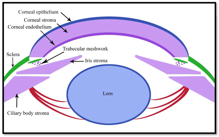
Development of the eye is closely associated with neural crest cell migration and specification. Eye development is extremely complex, as it requires the working of a combination of local factors, receptors, inductors, and signaling interactions between tissues such as the optic cup and periocular mesenchyme (POM). The POM is comprised of neural crest-derived mesenchymal progenitor cells that give rise to numerous important ocular structures including those tissues that form the optic cup and anterior segment of the eye. A number of genes are involved in the migration and specification of the POM such as PITX2, PITX3, FOXC1, FOXE3, PAX6, LMX1B, GPR48, TFAP2A, and TFAP2B. In this review, we will discuss the relevance of these genes in the development of the POM and how mutations and defects result in rare ocular diseases.
Keywords: animal models; anterior segment dysgenesis; development; genetics; neural crest; ocular disease.
© 2018 Wiley Periodicals, Inc.
Conflict of interest statement
The authors declare no conflict of interest.
Figures
References
-
- Akula M, Martino VB, Williams T, West-Mays J. Abnormal development and differentiation of the periocular mesenchyme in AP-2β neural crest cell knockout mice. Paper presented at the Association for Research in Vision and Ophthalmology; Baltimore, USA. 2017. May 8,
-
- Bassett EA, Pontoriero GF, Feng W, Marquardt T, Fini ME, Williams T, West-Mays J. Conditional deletion of activating protein-2α (AP-2α) in the developing retina demonstrates non-cell-autonomous roles for AP-2α in optic cup development. Molecular and Cellular Biology. 2007;27(21):7497–7510. - PMC - PubMed
Publication types
MeSH terms
Substances
Grants and funding
LinkOut - more resources
Full Text Sources
Other Literature Sources
Medical
