Review
. 2019 Mar 1;9(3):a033688.
doi: 10.1101/cshperspect.a033688.

Metastases in Prostate Cancer

Affiliations
Review

Metastases in Prostate Cancer

Federico La Manna et al. Cold Spring Harb Perspect Med. .

Erratum in

  • Erratum: Metastases in Prostate Cancer.
    La Manna F, Karkampouna S, Zoni E, De Menna M, Hensel J, Thalmann GN, Kruithof-de Julio M. La Manna F, et al. Cold Spring Harb Perspect Med. 2018 Jul 2;8(7):a035568. doi: 10.1101/cshperspect.a035568. Cold Spring Harb Perspect Med. 2018. PMID: 29967098 Free PMC article. No abstract available.

Abstract

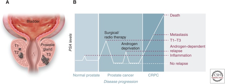
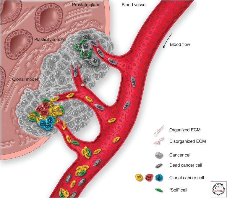
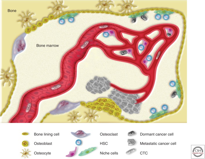
Prostate cancer (PCa) prognosis and clinical outcome is directly dependent on metastatic occurrence. The bone microenvironment is a favorable metastatic niche. Different biological processes have been suggested to contribute to the osteotropism of PCa such as hemodynamics, bone-specific signaling interactions, and the "seed and soil" hypothesis. However, prevalence of disseminating tumor cells in the bone is not proportional to the actual occurrence of metastases, as not all patients will develop bone metastases. The fate and tumor-reforming ability of a metastatic cell is greatly influenced by the microenvironment. In this review, the molecular mechanisms of bone and soft-tissue metastasis in PCa are discussed. Specific attention is dedicated to the residual disease, novel approaches, and animal models used in oncological translational research are illustrated.


Figures

Figure 1.
Figure 1.
Overview of prostate cancer (PCa) progression and therapeutic options. (A) Schematic drawing of different primary PCa stages, as defined by T category of the TNM staging system: T1, confined, not palpable tumor; T2, confined, palpable tumor; T3, palpable tumor, grown through the prostate capsule and spreading to the neighboring tissues. (B) Diagram of prostate serum antigen (PSA) blood levels over cancer progression. PSA serum level is used as a diagnostic marker to monitor both the progression of the disease and the effectiveness of the treatments received by the patient. CRPC, castrate-resistant prostate cancer.
Figure 2.
Figure 2.
The metastatic spread of prostate cancer (PCa) from the primary site. The primary lesion growth is characterized by a dysregulation of the prostate architecture, inducing changes in the extracellular matrix (ECM) composition and architecture, together with the induction of an inflammatory state that activates stromal cells and favors the recruitment of blood vessels to the lesion. As the tumor recruits new blood vessels, the systemic dissemination of cancer cells (in the form of circulating tumor cells, or CTCs) can take place. Two are the main models that explain the metastatic spread: “plasticity model” and “clonal model.” According to the plasticity model, as the cancer cells progress through malignancy, they may collect hits that make them fit for the metastatic process. The clonal model, on the other hand, theorizes that within the heterogeneous cancer cell subpopulations, clones with different fitness are generated, including some with the characteristics required for the metastatic spread. In addition, clusters of cells may form between spreading cancer and stromal cells from the primary lesion, the latter forming the “soil” cells that can facilitate the spreading and the survival of CTC. However, most of the CTCs will not survive in the bloodstream and will circulate as dead CTCs until clearance.
Figure 3.
Figure 3.
Dissemination of prostate cancer (PCa) cells in the bone marrow: from DTCs to overt metastasis. The hematopoietic bone tissue consists of two main parts: the bone tissue and the bone marrow. Bone is a highly regulated tissue that undergoes constant remodeling to keep its architecture and mechanical properties; as osteoclasts resorb weak bone, skeletal stem cells undergo local expansion, followed by differentiation into osteoblasts. Depending on the local microenvironment and the tissue architecture, osteoblasts will further differentiate either into bone-producing osteocytes or bone-lining cells—namely, the layer of cells that is in contact with the marrow cavities where hematopoiesis occurs. To ensure the lifelong production of blood, hematopoietic stem cells (HSCs) reside in specific areas, or niches, supported by specialized stromal cells (or niche cells) and can be found both at the endosteal and perivascular sides of the bone marrow. Within these niches, HSCs are induced in a state of quiescence, protected from cellular stresses, and prevented from further proliferation. Prostate CTCs may disseminate to the bone marrow and compete with the HSCs for the space in the niches. Within the niches, DTCs could remain dormant for an indefinite amount of time. Eventually, DTCs may exit their dormant state and start proliferating, bending the coupled processes of bone resorption and bone formation to support their growth. Most frequently, this vicious cycle produces hyperplastic bone tissue, eventually forming clinically relevant osteosclerotic metastasis.

References

    1. Adsan Ö, Cecchini MG, Bisoffi M, Wetterwald A, Klima I, Danuser HJ, Studer UE, Thalmann GN. 2002. Can the reverse transcriptase-polymerase chain reaction for prostate specific antigen and prostate specific membrane antigen improve staging and predict biochemical recurrence? BJU Int 90: 579–585. - PubMed
    1. Aguirre-Ghiso JA. 2007. Models, mechanisms and clinical evidence for cancer dormancy. Nat Rev Cancer 7: 834–846. - PMC - PubMed
    1. Arai F, Hirao A, Ohmura M, Sato H, Matsuoka S, Takubo K, Ito K, Koh GY, Suda T. 2004. Tie2/Angiopoietin-1 signaling regulates hematopoietic stem cell quiescence in the bone marrow niche. Cell 118: 149–161. - PubMed
    1. Arnold RS, Fedewa SA, Goodman M, Osunkoya AO, Kissick HT, Morrissey C, True LD, Petros JA. 2015. Bone metastasis in prostate cancer: Recurring mitochondrial DNA mutation reveals selective pressure exerted by the bone microenvironment. Bone 78: 81–86. - PMC - PubMed
    1. Batson OV. 1940. The function of the vertebral veins and their rôle in the spread of metastases. Ann Surgery 112: 138–149. - PMC - PubMed

LinkOut - more resources