. 2018 May 25;62(6):e00146-18.
doi: 10.1128/AAC.00146-18. Print 2018 Jun.

Triclosan Is an Aminoglycoside Adjuvant for Eradication of Pseudomonas aeruginosa Biofilms

Affiliations

Triclosan Is an Aminoglycoside Adjuvant for Eradication of Pseudomonas aeruginosa Biofilms

Michael M Maiden et al. Antimicrob Agents Chemother. .

Abstract

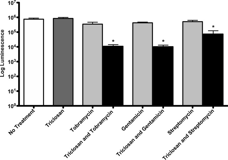
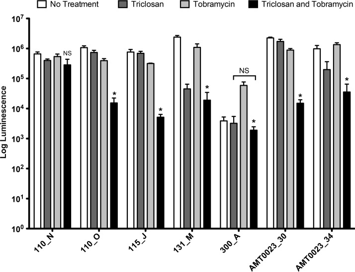
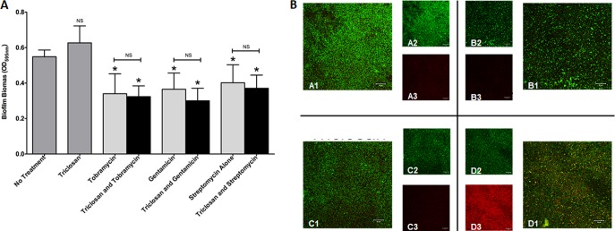
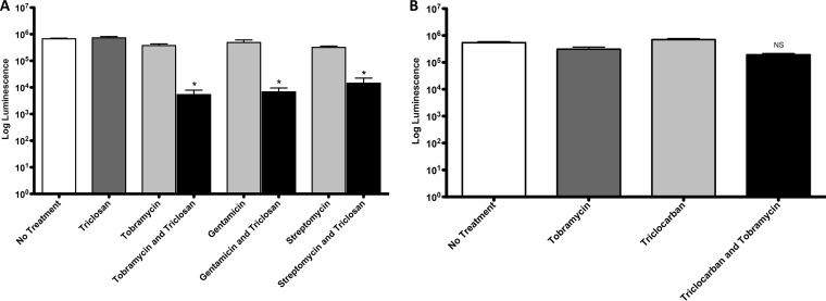
One of the most important clinical obstacles in cystic fibrosis (CF) treatment is antibiotic treatment failure due to biofilms produced by Pseudomonas aeruginosa The ability of this pathogen to survive eradication by tobramycin and pathoadapt into a hyperbiofilm state leading to chronic infections is key to its success. Retrospective studies have demonstrated that preventing this pathoadaptation by improving eradication is essential to extend the lives of CF patients. To identify adjuvants that enhance tobramycin eradication of P. aeruginosa, we performed a high-throughput screen of 6,080 compounds from four drug-repurposing libraries. We identified that the Food and Drug Administration (FDA)-approved compound triclosan, in combination with tobramycin, resulted in a 100-fold reduction of viable cells within biofilms at 6 h, but neither compound alone had significant antimicrobial activity against biofilms. This synergistic treatment significantly accelerated the killing of biofilms compared to that with tobramycin treatment alone, and the combination was effective against 6/7 CF clinical isolates compared to tobramycin treatment alone, including a tobramycin-resistant strain. Further, triclosan and tobramycin killed persister cells, causing a 100-fold reduction by 8 h and complete eradication by 24 h. Triclosan also enhances tobramycin killing of multiple Burkholderia cenocepacia and Staphylococcus aureus clinical isolates grown as biofilms. Additionally, triclosan showed synergy with other aminoglycosides, such as gentamicin or streptomycin. Triclosan is a well-tolerated aminoglycoside adjuvant shown to be safe for human use that could improve the treatment of biofilm-based infections.

Keywords: Pseudomonas aeruginosa; biofilm; persister; tobramycin; triclosan.


Figures

FIG 1
FIG 1
Molecular structure of triclosan and type II fatty acid synthesis. (A) Chemical structure of triclosan, 2,4,4′-trichloro-2′-hydroxydiphenyl ether. (B) Type II fatty acid synthesis (FASII) pathway. Triclosan is known to disrupt FASII by inhibiting FabI. However, FabV is more resistant to triclosan. ACP, acyl carrier protein; CoA, coenzyme A.
FIG 2
FIG 2
Triclosan enhances aminoglycoside killing of 24-h-old biofilms. Twenty-four-hour-old biofilms grown on minimum biofilm eradication concentration (MBEC) plates were treated for 6 h with 100 μM triclosan, 500 μM tobramycin, 100 μM gentamicin, or streptomycin, alone and in combination, and the number of viable cells within the biofilms was quantified using the BacTiter-Glo assay. The assay was performed at least three times in triplicate. The results represent the means ± the standard error of the mean (SEM). A one-way analysis of variance (ANOVA) followed by Bonferroni's multiple comparison post hoc test was used to determine statistical significance compared to each aminoglycoside alone (*, P < 0.05).
FIG 3
FIG 3
Triclosan enhances tobramycin killing at multiple concentrations. Twenty-four-hour-old biofilms grown on MBEC plates were treated for 6 h with 2-fold dilutions of equal concentrations of triclosan, tobramycin, and triclosan combined with tobramycin, and the number of viable cells within the biofilms was quantified using the BacTiter-Glo assay. The assay was performed at least three times in triplicate. The results represent the means ± the SEM.
FIG 4
FIG 4
Triclosan enhances low concentrations of tobramycin. Twenty-four-hour-old biofilms grown on MBEC plates were treated for 6 h with checkerboard dilutions of triclosan combined with tobramycin. Numbers of viable cells within the biofilms were quantified using the BacTiter-Glo assay. The assay was performed at least three times in triplicate, and the mean is shown.
FIG 5
FIG 5
Triclosan enhances the onset and maximum efficacy of tobramycin. Twenty-four-hour-old biofilms grown on MBEC plates were treated with triclosan (100 μM) tobramycin (500 μM), or a combination of the two. At 0, 2, 4, 6, and 8 h, the numbers of viable cells within the biofilms were determined using the BacTiter-Glo assay. The assay was performed at least three times in triplicate. The results represent means ± the SEM.
FIG 6
FIG 6
Tobramycin and triclosan are effective against P. aeruginosa CF isolates. Twenty-four-hour-old biofilms grown on MBEC plates were treated with triclosan (100 μM,) tobramycin (500 μM), or a combination of the two for 6 h. The numbers of viable cells within the biofilms were quantified using the BacTiter-Glo assay. The assay was performed at least three times in triplicate. The results represent means ± the SEM. A one-way ANOVA followed by Bonferroni's multiple comparison post hoc test was used to determine statistical significance compared to tobramycin alone (*, P < 0.05). NS, not significant.
FIG 7
FIG 7
Aminoglycosides combined with triclosan do not increase biofilm dispersal. (A) Twenty-four-hour-old biofilms grown on MBEC plates were treated with triclosan (100 μM), tobramycin (500 μM), gentamicin (100 μM), or streptomycin (100 μM), alone and in combination. The effect on biofilm biomass was quantified by staining with crystal violet. The experiment was performed at least five times in triplicate. The results represent the means ± SEM. A one-way ANOVA followed by Dunnett's multiple-comparison post hoc test was used to determine statistical significance compared to no treatment (*, P < 0.05). NS, not significant. (B) Twenty-four-hour-old biofilms grown in flow cells were treated with triclosan (100 μM), tobramycin (524 μM), or the combination for 6 h. Live cells are stained green, and dead cells are stained red. Representative images are shown: no treatment (top left quadrant), tobramycin alone (top right quadrant), triclosan alone (lower left quadrant), and a combination (lower right quadrant). Insets are shown for the live channel (A2, B2, C2, and D2) and for the dead channel (A3, B3, C3, and D3) for each condition.
FIG 8
FIG 8
FabI inhibition by triclosan is not responsible for the synergy. (A) Twenty-four-hour-old biofilms grown on MBEC plates by a FabI P. aeruginosa-deficient strain (Tn::fabI mutant) were treated for 6 h with triclosan (100 μM), tobramycin (500 μM), gentamicin (100 μM), or streptomycin (100 μM), alone and in combination. (B) Twenty-four-hour-old biofilms grown on MBEC plates by PAO1 were treated for 6 h with triclocarban (100 μM) or tobramycin (500 μM), alone or in combination. The number of viable cells within the biofilms were quantified using the BacTiter-Glo assay. The assay was performed at least three times in triplicate. The results represent means ± the SEM. A one-way ANOVA followed by Bonferroni's multiple-comparison post hoc test was used to determine statistical significance compared to tobramycin alone (*, P < 0.05). NS, not significant.
FIG 9
FIG 9
Tobramycin combined with triclosan kills persister cells. Twenty-hour-old stationary-phase cells were treated with triclosan (100 μM) or tobramycin (50 μM), alone and in combination, for 6 h. At 0, 2, 4, 6, 8, and 24 h, aliquots were taken for enumeration (CFU per milliliter). The experiment was performed three times in triplicate. The results represent means ± the SEM.

References

    1. O'Sullivan BP, Freedman SD. 2009. Cystic fibrosis. Lancet 373:1891–1904. doi:10.1016/S0140-6736(09)60327-5. - DOI - PubMed
    1. van Gool K, Norman R, Delatycki MB, Hall J, Massie J. 2013. Understanding the costs of care for cystic fibrosis: an analysis by age and health status. Value Health 16:345–355. doi:10.1016/j.jval.2012.12.003. - DOI - PubMed
    1. Ouyang L, Grosse SD, Amendah DD, Schechter MS. 2009. Healthcare expenditures for privately insured people with cystic fibrosis. Pediatr Pulmonol 44:989–996. doi:10.1002/ppul.21090. - DOI - PubMed
    1. Agrawal A, Agarwal A, Mehta D, Sikachi RR, Du D, Wang J. 2017. Nationwide trends of hospitalizations for cystic fibrosis in the United States from 2003 to 2013. Intractable Rare Dis Res 6:191–198. doi:10.5582/irdr.2017.01043. - DOI - PMC - PubMed
    1. Cutting GR. 2015. Cystic fibrosis genetics: from molecular understanding to clinical application. Nat Rev Genet 16:45–56. doi:10.1038/nrg3849. - DOI - PMC - PubMed

Publication types

MeSH terms