. 2018 Jun 21;86(7):e00706-17.
doi: 10.1128/IAI.00706-17. Print 2018 Jul.

Interleukin-22 Immunotherapy during Severe Influenza Enhances Lung Tissue Integrity and Reduces Secondary Bacterial Systemic Invasion

Affiliations

Interleukin-22 Immunotherapy during Severe Influenza Enhances Lung Tissue Integrity and Reduces Secondary Bacterial Systemic Invasion

Adeline Barthelemy et al. Infect Immun. .

Abstract

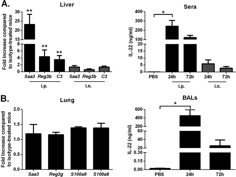
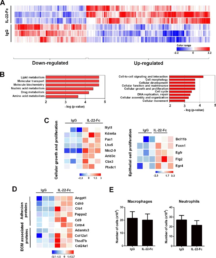
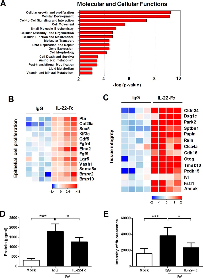
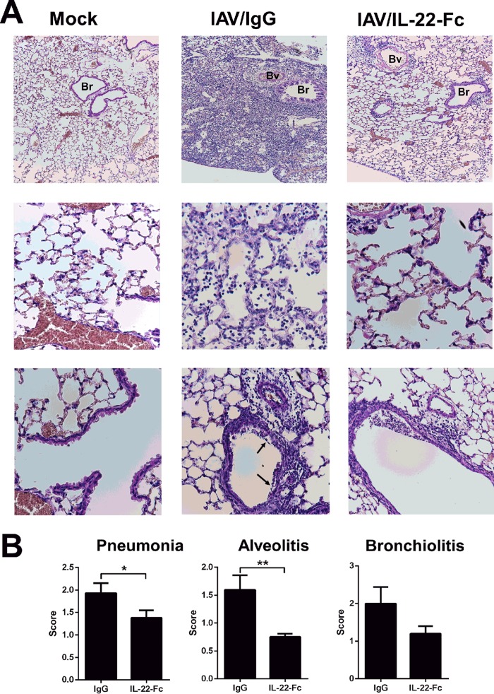
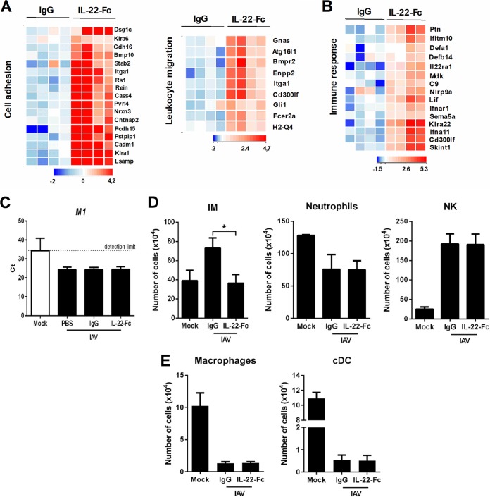
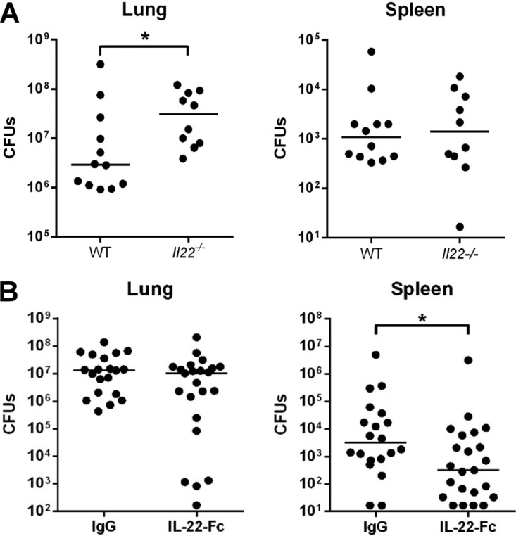
Severe bacterial (pneumococcal) infections are commonly associated with influenza and are significant contributors to the excess morbidity and mortality of influenza. Disruption of lung tissue integrity during influenza participates in bacterial pulmonary colonization and dissemination out of the lungs. Interleukin-22 (IL-22) has gained considerable interest in anti-inflammatory and anti-infection immunotherapy over the last decade. In the current study, we investigated the effect of exogenous IL-22 delivery on the outcome of pneumococcal superinfection postinfluenza. Our data show that exogenous treatment of influenza virus-infected mice with recombinant IL-22 reduces bacterial dissemination out of the lungs but is without effect on pulmonary bacterial burden. Reduced systemic bacterial dissemination was linked to reinforced pulmonary barrier functions, as revealed by total protein measurement in the bronchoalveolar fluids, intratracheal fluorescein isothiocyanate-dextran tracking, and histological approaches. We describe an IL-22-specific gene signature in the lung tissue of influenza A virus (IAV)-infected (and naive) mice that might explain the observed effects. Indeed, exogenous IL-22 modulates the gene expression profile in a way that suggests reinforcement of tissue integrity. Our results open the way to alternative approaches for limiting postinfluenza bacterial superinfection, particularly, systemic bacterial invasion.

Keywords: epithelial barrier; influenza; interleukin 22; superinfection.


Figures

FIG 1
FIG 1
Effect of IL-22 treatment on gene expression in the liver and in the lungs. (A) Mice were i.p. treated with 100 μg of IL-22-Fc or isotype control. After 24 h, liver RNAs were extracted, and gene expression was analyzed by quantitative RT-PCR. The concentration of IL-22 in the blood was measured by enzyme-linked immunosorbent assay at 24 h and 72 h after inoculation. (B) Mice were i.n. treated with 100 μg of IL-22-Fc or an isotype control. Lung RNAs were extracted at 24 h after treatment. IL-22 was measured by enzyme-linked immunosorbent assay in the BAL fluids at 24 h and 72 h postinoculation. Data are expressed as fold increase over average gene expression in isotype-treated mice and represent the means ± standard deviations. One representative experiment out of two (n = 3 to 6), except for panel A (n = 8, two pooled experiments), is shown. *, P < 0.05; **, P < 0.01 (Mann-Whitney t test for data in the left panels and a Kruskal-Wallis one-way ANOVA for data in the right panels).
FIG 2
FIG 2
Transcriptomic signature of IL-22 in the lungs of naive mice. (A) Total RNAs from Ig-treated mice or IL-22-Fc-treated mice (5 μg/animal) were extracted from the lungs at 8 h poststimulation. Hierarchical clustering diagrams showing individual replicates are represented. Normalized intensity values (IL-22-Fc condition compared to IgG condition) are depicted in blue (downregulated) or red (upregulated) (P < 0.05 and fold change of >2). The magnitude of the regulation is illustrated by the intensity of the color. Hierarchical clustering representation indicates a minimal effect of the isotype control in the lung tissue (data not shown). (B) Gene ontological analysis. Significantly enriched pathways are represented as a bar plot (IPA). For panels A and B, data from three mice are shown. (C) Heat maps of genes that were differentially regulated by IL-22 treatment and that belong to the cellular growth and proliferation family (GO:0008283) and, within this family, belonging to the epithelial growth and proliferation subfamily (GO:0050673). Representative genes are noted on the right. Each column represents data from one individual mouse, with three mice per group. (D) Heat maps of genes represented in the families of adhesion proteins (GO:0022610) and extracellular-matrix (ECM)-associated proteins (GO:0031012), both belonging to the morphology family. (E) Naive mice were treated with IL-22-Fc or IgG (5 μg/animal), and 8 h later, cells from whole lungs were analyzed by flow cytometry. The mean number ± standard deviation of alveolar macrophages (CD45+ Siglec F+ CD11blow) and neutrophils (CD11b+ Ly6G+ Siglec F) are depicted (n = 8; two independent experiments).
FIG 3
FIG 3
Effect of IL-22 supplementation on pulmonary gene expression and barrier functions. (A to C) Transcriptomic signature of IL-22 in the lungs of IAV-infected mice. Mice were i.n. infected with 500 PFU of the H1N1 pandemic IAV strain. At 7 days postinfection, mice were treated with PBS, the isotype control (5 μg/animal), or IL-22-Fc (5 μg/animal). Lung samples were collected 8 h later for further transcriptomic analysis. Ontological analysis is shown in panel A. Significantly enriched pathways are represented as a bar plot (IPA). Data from four mice are shown. In panels B and C, heat maps show genes that were differentially regulated by IL-22 treatment and that belong to the epithelial growth and proliferation family (GO:0050673) and to the tissue integrity family. Representative genes are noted on the right. Each column represents data from one individual mouse, with four mice per group. (D) Protein concentrations in the BAL fluids of IAV-infected mice treated with IL-22-Fc or IgG. Data represent the means ± standard deviations (n = 10 to 12; three pooled experiments). (E) IAV-infected mice, untreated or treated with IL-22, were inoculated with FITC-dextran. One hour later, FITC-dextran was quantified in the blood. Data represent the means ± standard deviations (n = 8; two pooled experiments). *, P < 0.05; ***, P < 0.001 (Kruskal-Wallis one-way ANOVA).
FIG 4
FIG 4
Histological analysis of lung sections from IAV-infected (at 7 days postinfection) mice untreated or treated with IL-22. (A) Representative lung sections stained with hematoxylin and eosin are shown. Alveolar lesions (middle row) and perivascular and peribronchic infiltrates combined with lesions of alveolitis (bottom row) are shown. Arrows indicate denuded epithelia. Bv, blood vessel, Br, bronchiole. (B) Sections were scored blindly for levels of immunopathology. Data represent the means ± standard deviations (n = 5 mice/group). *, P < 0.05; **, P < 0.01 (Mann-Whitney t test).
FIG 5
FIG 5
(A) Heat maps of genes belonging to cell adhesion (GO:0007155) and leukocyte migration (GO:0050900) families. (B) Heat map of genes belonging to the immune response family (GO:0006955). (C) Determination of the viral load after IL-22 treatment. IAV M1 mRNA levels were measured by quantitative RT-PCR. Data are expressed as cycle threshold (CT) values. The dashed line represents the detection threshold (n = 6). (D) IAV-infected mice were treated at 7 days postinfection with IL-22-Fc (5 μg/animal) or the isotype control. Eight hours later, cells from whole lungs were analyzed by flow cytometry. The mean number ± standard deviation of inflammatory monocytes (IM) (CD45+ Siglec F Ly6G Ly6C+ CD11b+ CCR2+), neutrophils (CD45+ CD11b+ Ly6G+ Siglec F), and NK cells (CD45+ NKp46+ TCRβ; TCRβ is T cell receptor beta) are depicted. (E) The mean numbers ± standard deviations of alveolar macrophages (CD45+ Siglec F+ CD11blow) and conventional dendritic cells (cDC) (CD45+ CD11chigh MHC class II+ Siglec F CD64; MHC is major histocompatibility complex) are depicted. (D and E) As a control, the number of immune cells from mock-treated mice are indicated (n = 8; two pooled experiments). The statistical analysis between mock-treated and IAV-infected mice is not depicted. *, P < 0.05 (Kruskal-Wallis one-way ANOVA).
FIG 6
FIG 6
Effect of endogenous and exogenous IL-22 in local and systemic bacterial loads in the context of influenza. (A) Role of endogenous IL-22 on secondary bacterial infection postinfluenza. Wild type (WT) or Il22−/− mice were infected with 500 PFU of the H1N1 pandemic IAV strain. Seven days later, IAV-infected mice were challenged with S. pneumoniae (1 × 10³ CFU). (B) Effect of IL-22 supplementation on bacterial superinfection postinfluenza. At day 7 post-IAV infection, mice were i.n. inoculated with 5 μg of IL-22-Fc or an isotype control. After 16 h, mice were challenged with S. pneumoniae. (A and B) Doubly infected mice were sacrificed 30 h after S. pneumoniae challenge, and the number of CFU was determined in the lungs and spleens. Total CFU counts in each tissue are represented. The solid line corresponds to the median values. Results from a pool of two independent experiments (n = 10 to 12 mice) (A) or three independent experiments (n = 20 to 23 mice) (B) are shown. *, P < 0.05 (Mann-Whitney t test).

References

    1. Morens DM, Taubenberger JK, Fauci AS. 2008. Predominant role of bacterial pneumonia as a cause of death in pandemic influenza: implications for pandemic influenza preparedness. J Infect Dis 198:962–970. doi:10.1086/591708. - DOI - PMC - PubMed
    1. McCullers JA. 2006. Insights into the interaction between influenza virus and pneumococcus. Clin Microbiol Rev 19:571–582. doi:10.1128/CMR.00058-05. - DOI - PMC - PubMed
    1. Snelgrove RJ, Godlee A, Hussell T. 2011. Airway immune homeostasis and implications for influenza-induced inflammation. Trends Immunol 32:328–334. doi:10.1016/j.it.2011.04.006. - DOI - PubMed
    1. Pittet LA, Hall-Stoodley L, Rutkowski MR, Harmsen AG. 2010. Influenza virus infection decreases tracheal mucociliary velocity and clearance of Streptococcus pneumoniae. Am J Respir Cell Mol Biol 42:450–460. doi:10.1165/rcmb.2007-0417OC. - DOI - PMC - PubMed
    1. Li N, Ren A, Wang X, Fan X, Zhao Y, Gao GF, Cleary P, Wang B. 2015. Influenza viral neuraminidase primes bacterial coinfection through TGF-β-mediated expression of host cell receptors. Proc Natl Acad Sci U S A 112:238–243. doi:10.1073/pnas.1414422112. - DOI - PMC - PubMed

Publication types