. 2018 Jun 22;293(25):9614-9628.
doi: 10.1074/jbc.M117.812016. Epub 2018 Apr 16.

Dual functionality of β-tryptase protomers as both proteases and cofactors in the active tetramer

Affiliations

Dual functionality of β-tryptase protomers as both proteases and cofactors in the active tetramer

Henry R Maun et al. J Biol Chem. .

Abstract

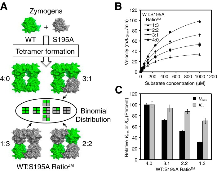
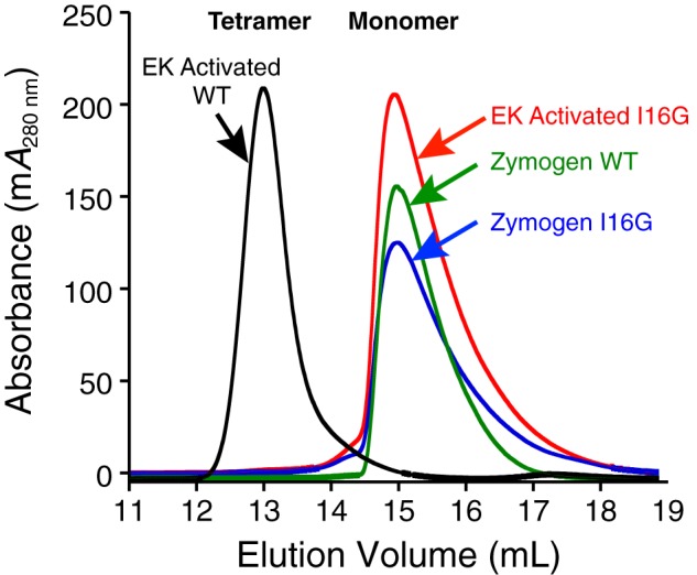
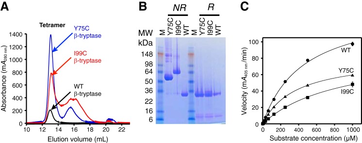
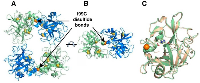
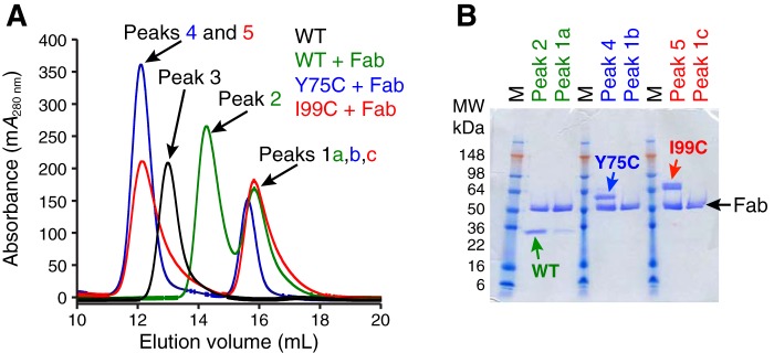
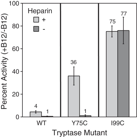
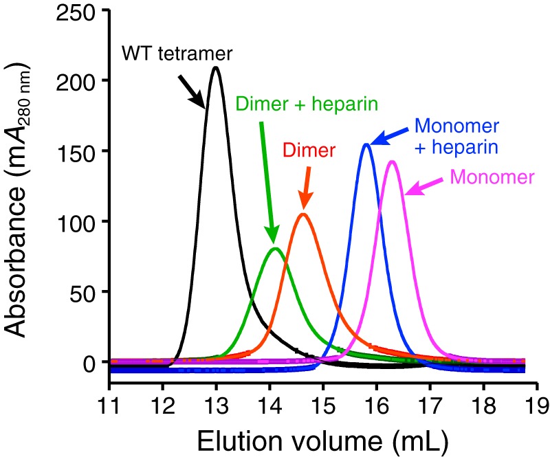
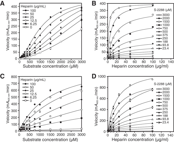
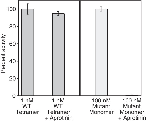
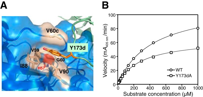
Human β-tryptase, a tetrameric trypsin-like serine protease, is an important mediator of the allergic inflammatory responses in asthma. During acute hypersensitivity reactions, mast cells degranulate, releasing active tetramer as a complex with proteoglycans. Extensive efforts have focused on developing therapeutic β-tryptase inhibitors, but its unique activation mechanism is less well-explored. Tryptase is active only after proteolytic removal of the pro-domain followed by tetramer formation via two distinct symmetry-related interfaces. We show that the cleaved I16G mutant cannot tetramerize, likely due to impaired insertion of its N terminus into its "activation pocket," indicating allosteric linkage at multiple sites on each protomer. We engineered cysteines into each of the two distinct interfaces (Y75C for small or I99C for large) to assess the activity of each tetramer and disulfide-locked dimer. Using size-exclusion chromatography and enzymatic assays, we demonstrate that the two large tetramer interfaces regulate enzymatic activity, elucidating the importance of this protein-protein interaction for allosteric regulation. Notably, the I99C large interface dimer is active, even in the absence of heparin. We show that a monomeric β-tryptase mutant (I99C*/Y75A/Y37bA, where C* is cysteinylated Cys-99) cannot form a dimer or tetramer, yet it is active but only in the presence of heparin. Thus heparin both stabilizes the tetramer and allosterically conditions the active site. We hypothesize that each β-tryptase protomer in the tetramer has two distinct roles, acting both as a protease and as a cofactor for its neighboring protomer, to allosterically regulate enzymatic activity, providing a rationale for direct correlation of tetramer stability with proteolytic activity.

Keywords: allosteric regulation; enzyme mechanism; heparin-binding protein; protein engineering; protein–protein interaction; serine protease; tetramerization; tryptase.


Conflict of interest statement

The authors were all employees of Genentech, Inc., when the work was carried out except for L. B. S. L. B. S. is a paid consultant for Genentech, Inc

Figures

Figure 1.
Figure 1.
Activity of tetrameric β-tryptase with different WT to S195A protomer ratios. A, cartoon depicting the generation of β-tryptase tetramers following enterokinase cleavage of WT and S195A zymogens at four different zymogen mixing ratios (RatioZM). The heterotetramers are actually a mixture of individual tetramer species weighted according to their binomial distribution (Table 1). B, comparison of the four β-tryptase tetramers with different protomer ratios at 1 nm measured with the chromogenic substrate S-2288. Data were collected in triplicate and fit to the Michaelis-Menten equation; errors are shown as S.D. C, comparison of Vmax and Km values of the different β-tryptase tetramer mixtures with WT; Vmax and Km were normalized to 100% for WT; errors are shown as S.D.
Figure 2.
Figure 2.
Size-exclusion chromatography of WT and I16G β-tryptase zymogens and proteases. WT β-tryptase forms tetramers after activation and pro-domain removal by EK in the presence of 0.5 mg/ml heparin. WT tetramer had an Ve of 13.0 ml on an S200GL column in SEC buffer. Following identical pro-domain removal by EK with heparin, I16G β-tryptase has a Ve of 15.0 ml in SEC buffer, which is essentially identical to that observed for zymogens of WT and I16G β-tryptase.
Figure 3.
Figure 3.
Disulfide engineering of β-tryptase dimers and tetramers. Tetrameric β-tryptase was engineered such that two of the four protomer interfaces were covalently linked by a disulfide bond. PyMOL was used to measure the distances between the two proposed thiols of Y75C in the small protomer interfaces (A:B and C:D) or I99C in the large protomer interfaces (A:D and B:C). Tetrameric β-tryptase having a mutation at either Y75C or I99C could only dissociate into two distinct disulfide-linked dimers as indicated in the cartoon. The red spheres are the oxygen atoms of Tyr-75. Protomer nomenclature corresponds to that described by Pereira et al. (15).
Figure 4.
Figure 4.
Characterization of WT, Y75C, and I99C β-tryptases. A, size-exclusion chromatography of tetrameric WT, Y75C, and I99C β-tryptases was carried out as in Fig. 2 on an S200GL column in SEC buffer; the dimeric zymogens of Y75C and I99C β-tryptases were activated and then run on SEC. Elution volumes for the tetrameric peaks were 13.0 ml, which were pooled separately. B, SDS-PAGE of pooled tetrameric WT, Y75C, and I99C β-tryptases under nonreducing (NR) and reducing (R) conditions. C, enzymatic activity of tetrameric WT, Y75C, and I99C β-tryptases at 1 nm concentration using S-2288 as a substrate. Data were collected in triplicate and fit to the Michaelis-Menten equation; errors are shown as S.D.
Figure 5.
Figure 5.
Crystal structure of tetrameric I99C β-tryptase mutant at 2.7 Å resolution. A, I99C β-tryptase mutant is shown in green and blue, with internal disulfides in yellow and the I99C disulfide in orange. Cysteine residues are shown as spheres for the disulfides; sulfur atoms are yellow for internal disulfides and orange for I99C disulfides. B, tetramer was rotated ∼90°; only one of the disulfide-locked dimers is shown. C, I99C protomer is superposed with one from the WT structure (1A0L), which is shown in beige. The catalytic triad residues are shown as sticks.
Figure 6.
Figure 6.
Characterization of WT, Y75C, and I99C β-tryptases in complex with murine B12 Fab. A, size-exclusion chromatography of B12 Fab in complex with WT, Y75C, and I99C β-tryptases was carried out as in Fig. 2 on an S200GL column in SEC buffer. Chromatograms are colored as follows: WT tryptase tetramer is colored black (Peak 3; Ve = 13.0 ml); complexes of B12 Fab with WT are green (Peak 2; Ve = 14.3 ml), Y75C is blue (Peak 4; Ve = 12.1 ml), and I99C is red (Peak 5; Ve = 12.1 ml). Excess free B12 Fab in Peaks 1a,b,c all have a similar Ve = 15.7 ml. B, fractions of main peaks were analyzed by SDS-PAGE under nonreducing conditions.
Figure 7.
Figure 7.
Activity of tryptase WT and disulfide-locked dimer mutants after dissociation with B12 Fab. The percent activity remaining of WT, Y75C, and I99C tryptase mutants (1 nm) in the presence of B12 Fab (125 nm) compared with its absence was determined in the presence (light gray) and absence (dark gray) of 0.1 mg/ml heparin. Tryptase variants were incubated with B12 Fab for 15 min at room temperature prior to activity measurements. The percent activity for each tryptase variant in the absence of B12 Fab and the presence and absence of 0.1 mg/ml heparin is defined as 100%. Data were collected in triplicate for two independent determinations; errors are shown as S.D.
Figure 8.
Figure 8.
Size-exclusion chromatography of tetrameric WT, dimeric I99C/Y75A/Y37bA, and monomeric I99C*/Y75A/Y37bA β-tryptases in the presence or absence of heparin. After pro-domain removal by EK, proteins were analyzed on an S200GL column in either SEC buffer (without heparin) or in TNH buffer (with heparin). Elution volumes for WT tetramer without heparin (13.0 ml), I99C/Y75A/Y37bA dimer with heparin (14.1 ml), I99C/Y75A/Y37bA dimer without heparin (14.6 ml), I99C*/Y75A/Y37bA monomer with heparin (15.8 ml), and I99C*/Y75A/Y37bA monomer without heparin (16.3 ml) are as indicated in parentheses.
Figure 9.
Figure 9.
Dependence of velocity on substrate and heparin for activated dimeric I99C/Y75A/Y37bA and monomeric I99C*/Y75A/Y37bA β-tryptase mutants. A, activated I99C/Y75A/Y37bA dimer data for the dependence of velocity with 2 mm S-2288 substrate at different heparin concentrations are shown. Data were collected in triplicate and fit to a sigmoidal equation. B, activated I99C/Y75A/Y37bA dimer data for the dependence of velocity on heparin at different S-2288 substrate concentrations are shown. C, activated I99C*/Y75A/Y37bA monomer data for the dependence of velocity on S-2288 substrate at different heparin concentrations are shown. D, activated I99C*:Y75A:Y37bA monomer data for the dependence of velocity on heparin at different S-2288 substrate concentrations are shown. Data for B, C, and D were each collected in triplicate and fit to hyperbolic binding equations and the 1:1 binding isotherm for B and D or Michaelis-Menten equation for C. Errors in all data are shown as S.D. Tryptase dimer and monomer concentrations were 100 and 400 nm, respectively.
Figure 10.
Figure 10.
Aprotinin only inhibits monomeric β-tryptase. Relative enzymatic activity of tetrameric WT and monomeric mutant (I99C*/Y75A/Y37bA) β-tryptases in the presence or absence of 10 μm aprotinin is shown. Activities were determined in TNH buffer with 1 mm S-2288. Tetramer activity was assayed at 1 nm and normalized to 1 nm WT; monomer activity was assayed at 100 nm and normalized to 100 nm mutant monomer. Data were collected in triplicate; errors are shown as S.D.
Figure 11.
Figure 11.
Characterization of the Y173dA tetramer mutant. A, Y173d from one protomer in the large interface lies in a hydrophobic pocket of its neighboring protomer. It is sandwiched between Val-90 and Val-60c with surrounding Val-59, Gly-60, and Ile-88 hydrophobic residues; the 60s loop is relatively close to the catalytic His-57 (red). B, dependence of reaction velocity on S-2288 substrate for tetrameric WT β-tryptase and the Y173dA mutant. Data were collected in triplicate and fit to the Michaelis-Menten equation; errors are shown as S.D.

References

    1. Caughey G. H. (2016) Mast cell proteases as pharmacological targets. Eur. J. Pharmacol. 778, 44–55 10.1016/j.ejphar.2015.04.045 - DOI - PMC - PubMed
    1. Hallgren J., and Pejler G. (2006) Biology of mast cell tryptase. An inflammatory mediator. FEBS J. 273, 1871–1895 10.1111/j.1742-4658.2006.05211.x - DOI - PubMed
    1. McNeil H. P., Adachi R., and Stevens R. L. (2007) Mast cell-restricted tryptases: structure and function in inflammation and pathogen defense. J. Biol. Chem. 282, 20785–20789 10.1074/jbc.R700017200 - DOI - PubMed
    1. Wernersson S., and Pejler G. (2014) Mast cell secretory granules: armed for battle. Nat. Rev. Immunol. 14, 478–494 10.1038/nri3690 - DOI - PubMed
    1. Rivera J., Fierro N. A., Olivera A., and Suzuki R. (2008) New insights on mast cell activation via the high affinity receptor for IgE. Adv. Immunol. 98, 85–120 10.1016/S0065-2776(08)00403-3 - DOI - PMC - PubMed

Publication types