. 2018 Apr 16;9(1):1490.
doi: 10.1038/s41467-018-03746-3.

In silico optimization of a guava antimicrobial peptide enables combinatorial exploration for peptide design

Affiliations

In silico optimization of a guava antimicrobial peptide enables combinatorial exploration for peptide design

William F Porto et al. Nat Commun. .

Abstract

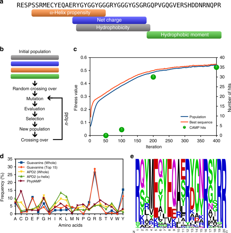
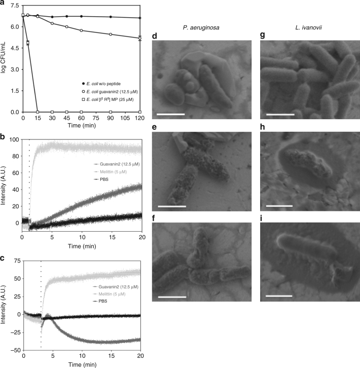
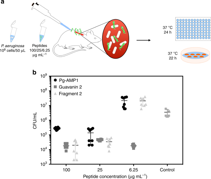
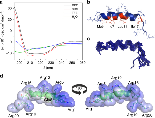
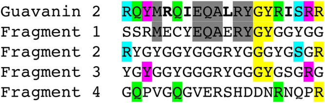
Plants are extensively used in traditional medicine, and several plant antimicrobial peptides have been described as potential alternatives to conventional antibiotics. However, after more than four decades of research no plant antimicrobial peptide is currently used for treating bacterial infections, due to their length, post-translational modifications or high dose requirement for a therapeutic effect . Here we report the design of antimicrobial peptides derived from a guava glycine-rich peptide using a genetic algorithm. This approach yields guavanin peptides, arginine-rich α-helical peptides that possess an unusual hydrophobic counterpart mainly composed of tyrosine residues. Guavanin 2 is characterized as a prototype peptide in terms of structure and activity. Nuclear magnetic resonance analysis indicates that the peptide adopts an α-helical structure in hydrophobic environments. Guavanin 2 is bactericidal at low concentrations, causing membrane disruption and triggering hyperpolarization. This computational approach for the exploration of natural products could be used to design effective peptide antibiotics.


Conflict of interest statement

The authors declare no competing interests.

Figures

Fig. 1
Fig. 1
Design and selection of guavanins. a Fragment mapping into the Pg-AMP1 sequence. Each fragment represents the maximum value of its respective physicochemical property: in orange, the α-helix propensity (0.553); in blue, the positive net charge (+3); in gray, the average hydrophobicity (−0.092); and in green, the hydrophobic moment (0.3). b Flowchart of our custom genetic algorithm. The four Pg-AMP1 fragments were used as the initial population; in the first iteration a totally random sequence selection system for crossing over was applied, in order to improve the diversity of sequences and in the subsequent iterations a roulette wheel selection model was applied for selection of sequences for crossing over. c Fitness function evolution during the algorithm iterations and the number of CAMP hits of the highest ranked sequence at iterations 50, 100, 200, and 400; Guavanin sequences were retrieved from the 50th iteration. d Amino acid distribution of guavanins and AMPs from APD2 and PhytAMP. Blue squares represent data obtained from 100 guavanin sequences; orange diamonds, the top 15 guavanins; yellow down triangles, the overall APD2 composition; green up triangles, the composition of α-helical peptides from APD2; and brown right triangles the plant AMP sequences from PhytAMP (37). e The frequency logo of the 100 generated guavanin sequences (Supplementary Table 1), showing that they are arginine-rich peptides, Arg residues are in at least 20% of their compositions
Fig. 2
Fig. 2
Killing and membrane effects of guavanin 2. a Time–kill profile of guavanin 2 against E. coli ATCC25922. Positive and negative controls correspond to bacteria incubated [I5, R8] mastoparan and without peptide, respectively. The data are the means ± S.E.M. of one experiments executed in triplicate. b, c Effect of guavanin 2 on plasma membrane integrity of E. coli ATCC 25922 cells after addition (vertical dotted line) of a concentration of peptide twofold above the MIC (12.5 µM). The pore-forming peptide melittin (5 µM) was used as a positive control. The negative control PBS corresponds to the bacteria incubated with the fluorescent probes without peptide. b Time-course cytoplasmic membrane permeation analysis of SYTOX Green uptake. cCytoplasmic membrane hyperpolarization using DiSC3(5). d SEM-FEG visualization of the effect of guavanin 2 on P. aeruginosa (df) and L. ivanovii (gi). The Controls without peptide are displayed in the d, g panels, respectively.  Bacteria were treated with a concentration of guavanin 2 corresponding to 25 µM (e), 50 µM (f, h) and 100 µM (i), respectively. Scale bar = 1 µm
Fig. 3
Fig. 3
In vivo activity of guavanin 2. a Schematic of the experimental design. Briefly, the back of mice was shaved and an abrasion was generated to damage the stratum corneum and the upper layer of the epidermis. Subsequently, an aliquot of 50 μL containing 5 × 107 CFU of P. aeruginosa in PBS was inoculated over each defined area. One day after the infection, peptides Pg-AMP1, guavanin 2, and Pg-AMP1 charge fragment were administered to the infected area. Animals were euthanized and the area of scarified skin was excised. b Four  days post-infection, homogenized using a bead beater for 20 min (25 Hz), and serially diluted for CFU quantification. Two independent experiments were performed with four mice per group in each case. Statistical significance was assessed using a two-way ANOVA. At all doses tested treatment with guavanin 2 significantly reduced CFU counts (p < 0.0001). Treatment with Pg-AMP1 and fragment 2 led to a significant reduction of bacterial load only at higher concentrations (25 and 100 µg mL−1)
Fig. 4
Fig. 4
Structure analysis of guavanin 2. a CD spectra of guavanin 2. The spectra in DPC (20 mM), SDS (20 mM) and TFE 50% were obtained at 25°C, pH 4 and a peptide concentration of 38 μM. The spectra in water was obtained at 25°C, pH 7 and a peptide concentration of 33 μM. Guavanin 2 has no defined structure in water, whereas it presents α-helical structure with hydrophobic solvents, in special DPC, which was selected for solving the three-dimensional structure by nuclear magnetic resonance (NMR). b Solution NMR structure of guavanin 2 in 100 mM (DPC-d38) micelles; a ribbon representation structure of lowest energy structure with side chains labeled (blue is hydrophilic and red is hydrophobic). c Ensemble of 15 backbone structures with low energy. d Electrostatic surfaces of guavanin 2 in 100 mM (DPC-d38) micelles. Surface potentials were set to ±5 kT e−1 (133.56 mV). Blue indicates positively charged regions and white apolar ones. Charged residues are labeled
Fig. 5
Fig. 5
Guavanin 2 and its ancestors. Guavanin 2 and the Pg-AMP1 fragments were aligned without including gaps to demonstrate the guavanin 2 inherited residues and mutations. The residues inherited from each the fragments are highlighted in gray (fragment 1—α-helix propensity), cyan (fragment 2—net charge), magenta (fragment 3—hydrophobicity), light green (fragment 4—hydrophobic moment), and yellow (two or more fragments); and the mutated residues are in bold face

References

    1. Peleg AY, Hooper DC. Hospital-acquired infections due to gram-negative bacteria. N. Engl. J. Med. 2010;362:1804–1813. doi: 10.1056/NEJMra0904124. - DOI - PMC - PubMed
    1. Gaynes R, Edwards JR. Overview of nosocomial infections caused by gram-negative bacilli. Clin. Infect. Dis. 2005;41:848–854. doi: 10.1086/432803. - DOI - PubMed
    1. Fleischmann C, et al. Assessment of global incidence and mortality of hospital-treated sepsis. Current estimates and limitations. Am. J. Respir. Crit. Care Med. 2016;193:259–272. doi: 10.1164/rccm.201504-0781OC. - DOI - PubMed
    1. Coates ARM, Halls G, Hu Y. Novel classes of antibiotics or more of the same? Br. J. Pharmacol. 2011;163:184–194. doi: 10.1111/j.1476-5381.2011.01250.x. - DOI - PMC - PubMed
    1. Cândido, E. S. et al. in Science Against Microbial Pathogens: Communicating Current Research and Technological Advances (ed. Méndez-Vilas, A.) 951–960 (Formatex, Badajoz, Spain, 2011).

Publication types

MeSH terms