. 2018 Apr 16;8(1):6034.
doi: 10.1038/s41598-018-24380-5.

HadD, a novel fatty acid synthase type II protein, is essential for alpha- and epoxy-mycolic acid biosynthesis and mycobacterial fitness

Affiliations

HadD, a novel fatty acid synthase type II protein, is essential for alpha- and epoxy-mycolic acid biosynthesis and mycobacterial fitness

Cyril Lefebvre et al. Sci Rep. .

Abstract

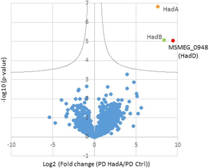
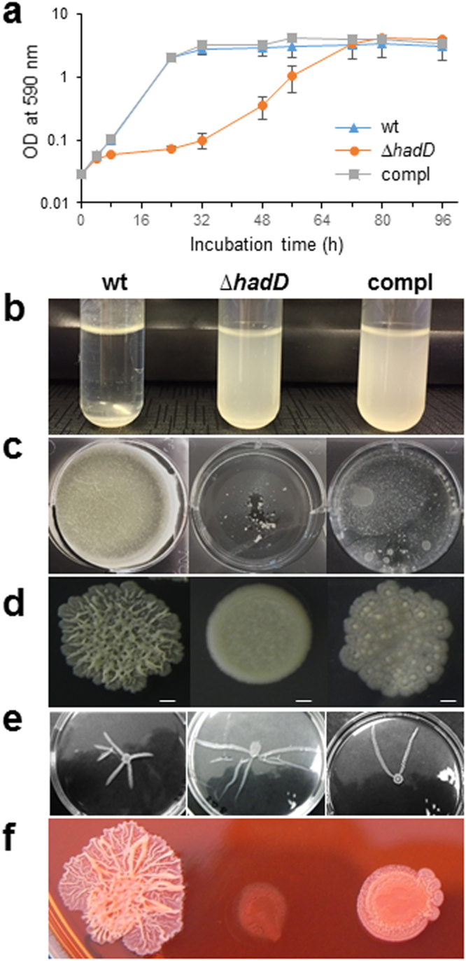
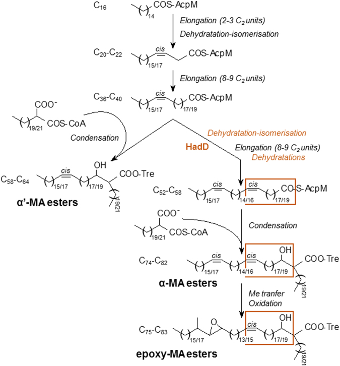
Mycolic acids (MAs) have a strategic location within the mycobacterial envelope, deeply influencing its architecture and permeability, and play a determinant role in the pathogenicity of mycobacteria. The fatty acid synthase type II (FAS-II) multienzyme system is involved in their biosynthesis. A combination of pull-downs and proteomics analyses led to the discovery of a mycobacterial protein, HadD, displaying highly specific interactions with the dehydratase HadAB of FAS-II. In vitro activity assays and homology modeling showed that HadD is, like HadAB, a hot dog folded (R)-specific hydratase/dehydratase. A hadD knockout mutant of Mycobacterium smegmatis produced only the medium-size alpha'-MAs. Data strongly suggest that HadD is involved in building the third meromycolic segment during the late FAS-II elongation cycles, leading to the synthesis of the full-size alpha- and epoxy-MAs. The change in the envelope composition induced by hadD inactivation strongly altered the bacterial fitness and capacities to aggregate, assemble into colonies or biofilms and spread by sliding motility, and conferred a hypersensitivity to the firstline antimycobacterial drug rifampicin. This showed that the cell surface properties and the envelope integrity were greatly affected. With the alarmingly increasing case number of nontuberculous mycobacterial diseases, HadD appears as an attractive target for drug development.


Conflict of interest statement

The authors declare no competing interests.

Figures

Figure 1
Figure 1
The dehydratase HadAB of FAS-II displays highly specific interaction with a novel protein, HadD (MSMEG_0948). Proteomics analysis of pull-down fractions using eGFP-HadA as a bait. Volcano plot [−log10 (p-value) versus log2 (fold change)] showing enriched proteins in eGFP-HadA-based pull-down (PD) versus control pull-down (using simple eGFP as a bait). Statistical analysis was performed from four independent biological replicates by two-sided Student t-test, variance correction and permutation-based false discovery rate (FDR) control in Perseus. Proteins considered as significantly enriched (FDR <5%, hyperbolic selection curve indicated in grey) are colored; the bait, HadA, is in orange, while copurifying proteins, HadB and HadD, are in green and red, respectively.
Figure 2
Figure 2
HadD belongs to the hydratase/dehydratase family. (a) Enzyme activity assays. Kinetic experiments were performed in 100 mM sodium phosphate buffer pH 7.0 in the presence of 10 µM C12:1-CoA and various concentrations of purified H-HadD protein, and monitored spectrophotometrically by following the disappearance of the trans-2-ethylenic function of the substrate at 263 nm. (b) MALDI-TOF MS analysis of the reaction product. Ions peaks of the hydration product, the 3-hydroxydodecanoyl-CoA, were detected in assays in the presence of HadD (bottom) but not in control assays without protein (top). The substrate ion peaks (proton and mono- to tetra-sodium adducts) are labeled in black and the hydration product ion peaks (mono- to tetra-sodium adducts) are labeled in red.
Figure 3
Figure 3
The lack of HadD profoundly changes the distribution and fine structure of the cell-wall-linked mycolic acids. (a) Structures of the three MA types from M. smegmatis. One of the main molecules was drawn for each type, as an example. The three segments of the meromycolic chain, delimited by the distal (D) and proximal (P) positions of the chemical functions, are indicated. (b) HPTLC profiles of cell-wall-linked MAMEs in the wt, ∆hadD, complemented (compl) and ∆pE strains. Five µg of each MA mixture were loaded onto a HPTLC plate developed in petroleum ether/diethyl ether (9:1, v/v), and stained by immersion in CuSO4 and heating. The figure is representative of four independent experiments. (c) MA distribution in each strain deduced from the quantification of the HPTLC band intensities as in panel (b). Data are means ± average deviations of four independent experiments. (d) MALDI-TOF MS spectra of the cell-wall-linked MAME mixtures. Peaks correspond to monosodium adducts. The major ion peaks are labelled with the matching MA type and the total carbon number (of the free acid form). ɛ, epoxy-MAMEs; their mass values are in italic. The spectra are representative of three independent experiments.
Figure 4
Figure 4
Alteration of the extractable lipid content upon hadD deletion. Thin layers are representative of at least three independent experiments. (a,b) TLC analyses of the total extractable lipids. Identical amounts of lipid mixtures were loaded on TLC plates developed either in CHCl3:CH3OH:H2O (65:25:4, v/v/v) (panel (a)) or in CHCl3:CH3OH (9:1, v/v) (panel (b)). Insert in panel (b): lower amounts of lipids were loaded to better visualize the Rf difference between TDM and TDM∆. For full picture, see Supplementary Figure S5c. The spots were revealed by anthrone spraying and heating. CL, cardiolipins; GPLs, glycopeptidolipids; PIMs, phosphatidylinositol mannosides; PG, phosphatidyl glycerol; TDM, trehalose dimycolate; TDM∆, TDMs of M. smegmatishadD; TMM, trehalose monomycolate; TMM∆, TMMs of M. smegmatishadD; TPPs, trehalose polyphleates; X, compound X. Compl, complemented strain; ∆pE, M. smegmatis pE deletion mutant lacking pE acyltranferase. The apolar GPLs are diglycosylated and the polar GPLs are triglycosylated. (c,d) MALDI-TOF MS spectra of purified TMMs and TDMs, respectively. Peaks correspond to monosodium adducts. For clarity, m/z values are indicated for the major ion peaks only. Peaks of TDM α + α are present in the wt strain but globally less intense than TDM α + ɛ. The type and total carbon number of MA chains are mentioned. Mass values of epoxy-MA containing lipids are in italic. ɛ, epoxy-MA chains.
Figure 5
Figure 5
HadD deletion has an impact on bacterial fitness, assembly, spreading and surface hydrophobicity. Comparison of M. smegmatis wt, ∆hadD and complemented (compl) strains in different phenotyping assays. All the pictures are representative of three independent experiments. (a) Planktonic growth. Curves were established at 37 °C in 7H9-based medium supplemented with 0.05% (w/v) Tween-80. The cfu numbers determined at 0, 24 and 48 h confirmed a significant growth reduction for M. smegmatis ΔhadD. Data are means and standard deviations of three independent experiments. (b) Aggregation assays. Samples of cultures grown until saturation in 7H9-based medium without Tween were kept unshaken. Photographs correspond to the 15 min time point of the assays (Supplementary Fig. S9a). (c) Biofilm formation at the air-liquid interface. It was monitored on Sauton’s medium after 6 days of incubation at 37 °C. (d) Colony morphology. Five µl culture aliquots were spotted on 7H10-based medium. Scale bars represent 1 mm. (e) Sliding motility. It was monitored after 7 day incubation on semi-solid 7H9-based medium. Finger-like extensions appeared and spread outwards from the central inoculation point. M. smegmatishadD displayed a higher number and much longer extensions than the wt strain, whereas the complemented strain exhibited an intermediate extension number. For sliding motility quantification, see Supplementary Fig. S9b. (f) Colony morphology on Congo red. Five µl preculture aliquots were spotted on 7H10-based medium supplemented with 100 µg/ml Congo red. For quantification of Congo red binding, see Supplementary Fig. S9c.
Figure 6
Figure 6
Increased sensitivity to low temperature, SDS and rifampicin upon hadD inactivation. Comparison of M. smegmatis wt, ∆hadD and complemented (compl) strains in different assays. Cultures were serially diluted then spotted on 7H10 medium supplemented with the required drug or detergent and incubated at 37 °C in standard conditions or at 30 °C and 42 °C for temperature testing. All the photographs are representative of three independent experiments. Susceptibility to (a) temperature, (b) SDS, (c) antimycobacterial drugs. The target protein of each drug is mentioned. RpoB is a DNA-directed RNA polymerase (β chain); InhA is the trans-2-enoyl-ACP reductase of FAS-II; EmbB is an arabinosyl transferase.
Figure 7
Figure 7
Proposed function of HadD in the mycolic acid biosynthesis pathway in M. smegmatis. The biosynthesis process of the meromycolic chain starts from the methyl-terminal end. After a first set of FAS-II elongation cycles, the possible dehydratation-isomerization of a C20-C22 3-hydroxyacyl-AcpM intermediate would allow the introduction of the future distal cis double bond (Fig. 3a) in the meromycolic chains. After a second set of FAS-II elongation cycles, a Claisen-type condensation of the resulting short α′-meromycoloyl chains with a carboxy-C22/C24 fatty acyl-CoA by Pks13 enzyme, followed by transfer onto trehalose (Tre) and reduction, generates the trehalose α′-MA esters. A second possible dehydratation-isomerization of the α′-meromycoloyl chains would introduce the future proximal cis double bond (Fig. 3a). After a third set of FAS-II elongation cycles including (3R)-hydroxyacyl-AcpM dehydratation steps, the condensation of the resulting long α-meromycoloyl chains with a carboxy-C22/C24 fatty acyl-CoA generates the trehalose α-MA esters. Finally, methyl transfer and oxidation reactions allow the biosynthesis of the epoxy-MA esters. HadD would catalyze either the dehydratation-isomerization or the dehydratation reactions during the synthesis of the third meromycolic segment (orange frame) (see also Fig. 3a). The total carbon number of the different acyl chains are mentioned.

References

    1. WHO. Global tuberculosis report 2017. (ed. WHO Press) (World Health Organization, 2017).
    1. Nishiuchi Y, Iwamoto T, Maruyama F. Infection sources of a common non-tuberculous mycobacterial pathogen, Mycobacterium avium complex. Front. Med. 2017;4:27. doi: 10.3389/fmed.2017.00027. - DOI - PMC - PubMed
    1. Brown-Elliott, B. A. & Philley, J. V. Rapidly growing mycobacteria. Microbiology spectrum5, TNMI7-0027–2016 (2017). - PMC - PubMed
    1. Sousa S, Bandeira M, Carvalho PA, Duarte A, Jordao L. Nontuberculous mycobacteria pathogenesis and biofilm assembly. Int. J. Mycobacteriol. 2015;4:36–43. doi: 10.1016/j.ijmyco.2014.11.065. - DOI - PubMed
    1. Soni I, De Groote MA, Dasgupta A, Chopra S. Challenges facing the drug discovery pipeline for non-tuberculous mycobacteria. J. Med. Microbiol. 2016;65:1–8. doi: 10.1099/jmm.0.000198. - DOI - PubMed

Publication types

MeSH terms