. 2018 May;24(5):610-616.
doi: 10.1038/s41591-018-0001-2. Epub 2018 Apr 16.

A single injection of crystallizable fragment domain-modified antibodies elicits durable protection from SHIV infection

Affiliations

A single injection of crystallizable fragment domain-modified antibodies elicits durable protection from SHIV infection

Rajeev Gautam et al. Nat Med. 2018 May.

Abstract

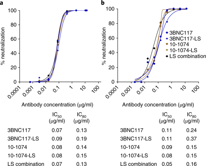
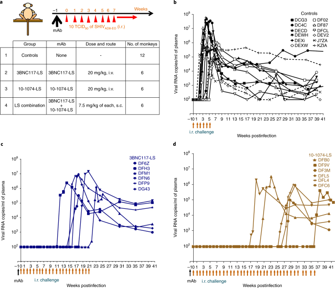
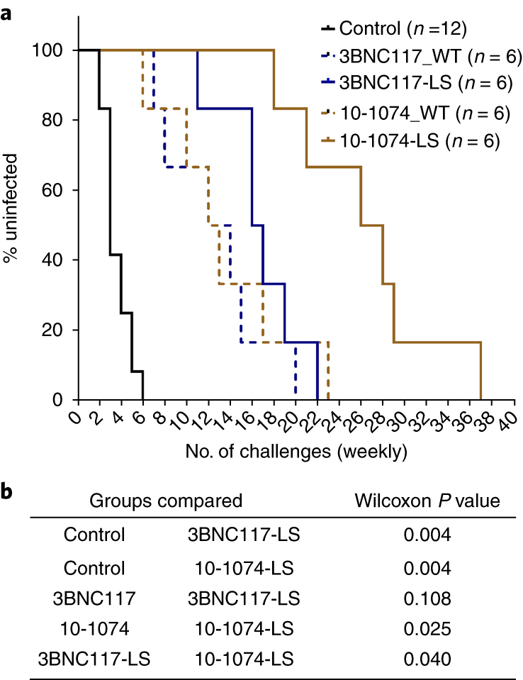
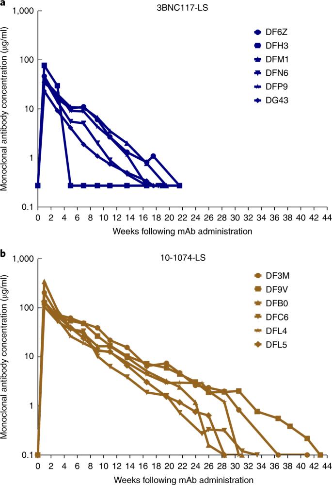
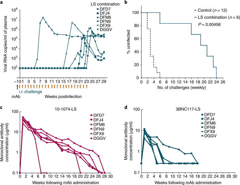
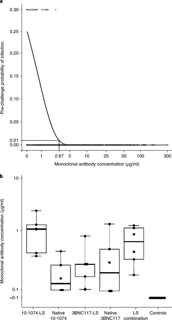
In the absence of an effective and safe vaccine against HIV-1, the administration of broadly neutralizing antibodies (bNAbs) represents a logical alternative approach to prevent virus transmission. Here, we introduced two mutations encoding amino acid substitutions (M428L and N434S, collectively referred to as 'LS') into the genes encoding the crystallizable fragment domains of the highly potent HIV-specific 3BNC117 and 10-1074 bNAbs to increase their half-lives and evaluated their efficacy in blocking infection following repeated low-dose mucosal challenges of rhesus macaques (Macaca mulatta) with the tier 2 SHIVAD8-EO. A single intravenous infusion of 10-1074-LS monoclonal antibodies markedly delayed virus acquisition for 18 to 37 weeks (median, 27 weeks), whereas the protective effect of the 3BNC117-LS bNAb was more modest (provided protection for 11-23 weeks; median, 17 weeks). Serum concentrations of the 10-1074-LS monoclonal antibody gradually declined and became undetectable in all recipients between weeks 26 and 41, whereas the 3BNC117-LS bNAb exhibited a shorter half-life. To model immunoprophylaxis against genetically diverse and/or neutralization-resistant HIV-1 strains, a combination of the 3BNC117-LS plus 10-1074-LS monoclonal antibodies was injected into macaques via the more clinically relevant subcutaneous route. Even though the administered mixture contained an amount of each bNAb that was nearly threefold less than the quantity of the single monoclonal antibody in the intravenous injections, the monoclonal antibody combination still protected macaques for a median of 20 weeks. The extended period of protection observed in macaques for the 3BNC117-LS plus 10-1074-LS combination could translate into an effective semiannual or annual immunoprophylaxis regimen for preventing HIV-1 infections in humans.


Conflict of interest statement

The authors declare no competing interests.

Figures

Fig. 1
Fig. 1. Neutralization sensitivity of broadly acting neutralizing anti-HIV-1 monoclonal antibodies against SHIVAD8-EO.
a, Top, neutralizing activity of the indicated bNAbs was determined against SHIVAD8-EO pseudovirions using TZM-bl target cells. Bottom, the calculated IC50 and IC80 values for the antibodies. b, Top, neutralizing activity of the indicated bNAbs was determined against replication-competent SHIVAD8-EO in a single-round TZM-bl infectivity assay in the presence of indinavir. Bottom, the calculated IC50 and IC80 values for the antibodies.  The neutralization assays were repeated three times with similar results.
Fig. 2
Fig. 2. Crystallizable fragment domain–modified HIV monoclonal antibodies confer durable protection against repeated low-dose IR SHIVAD8-EO challenges.
a, Experimental design for assessment of the protective efficacy of monoclonal antibodies in rhesus macaques. Single doses of the indicated individual monoclonal antibodies (20 mg per kg body weight) or combination monoclonal antibodies (7.5 mg per kg body weight of each monoclonal antibody) were administered either i.v. or s.c. Macaques were challenged with SHIVAD8-EO via the i.r. route weekly, beginning 1 week following monoclonal antibody (mAb) infusion. b, Plasma viral loads in rhesus macaques receiving no monoclonal antibody (controls; n = 12) challenged weekly with SHIVAD8-EO. c,d, Plasma viral loads in rhesus macaques (n = 6 per group) challenged weekly with SHIVAD8-EO beginning 1 week after i.v. administration of 3BNC117-LS (c) or 10-1074-LS (d) monoclonal antibody.
Fig. 3
Fig. 3. Protective effects of 3BNC117-LS and 10-1074-LS monoclonal antibodies against virus acquisition in rhesus macaques.
a, Kaplan–Meier analysis was used to assess infection rates for controls and recipients of 3BNC117-LS and 10-1074-LS monoclonal antibodies and their native forms. The percentage of uninfected rhesus macaques following SHIVAD8-EO i.r. challenge was assessed for the monoclonal antibody recipients (n = 6 per group) and control monkeys (n = 12). b, P values were determined using Wilcoxon rank-sum test (two-sided) comparing the number of challenges resulting in infections of control monkeys versus the individual monoclonal antibody–recipient group or between different monoclonal antibody–recipient groups.
Fig. 4
Fig. 4. Serum antibody concentrations in rhesus macaques infused with crystallizable fragment domain–modified monoclonal antibodies.
a, Concentrations of 3BNC117-LS antibody were measured in serum over the course of 6 months following a single i.v. infusion (20 mg per kg body weight) of the 3BNC117-LS monoclonal antibody using the TZM-bl cell assay. b, Concentrations of 10-1074-LS antibody were measured in serum for 9 months after i.v. infusion of a single 20 mg per kg body weight dose of 10-1074-LS monoclonal antibody using a TZM-bl cell assay. The assay was performed twice.
Fig. 5
Fig. 5. Protection efficacy of combination 3BNC117-LS plus 10-1074-LS monoclonal antibody administered s.c. to rhesus macaques.
a, Plasma viral loads in rhesus macaques (n = 6) challenged repeatedly with SHIVAD8-EO beginning 1 week after the s.c. administration of a single dose of the 3BNC117-LS plus 10-1074-LS monoclonal antibody mixture (7.5 mg per kg body weight of each). b, Kaplan–Meier survival curves show the percentage of rhesus macaques remaining uninfected following repeated SHIVAD8-EO i.r. challenges required to establish infection of monoclonal antibody–combination recipients (n = 6) or controls (n = 12). P values were determined using the Wilcoxon rank-sum test (two-sided) comparing the number of challenges resulting in infection of the controls and monoclonal antibody–combination recipients. c,d, Concentrations of 10-1074-LS and 3BNC117-LS monoclonal antibodies were measured, using the TZM-bl cell assay, in serum of rhesus macaques administered a single injection of the monoclonal antibody mixture s.c. Antibody concentrations were measured twice.
Fig. 6
Fig. 6. Antibody concentration predicts the probability of infection.
a, Probit regression was used to model the probability of infection based on antibody concentrations in serum of rhesus macaques at the time of each SHIVAD8-EO challenge. The probability of infection for the control monkeys (n = 12) was estimated to be 0.27. The fitted probability, determined using the probit regression model, is plotted for all recipients of LS-modified monoclonal antibodies (n = 18) and native monoclonal antibodies (n = 12; previously reported). Each red circle indicates the monoclonal antibody concentration at the time of virus challenge resulting in infection; blue circles indicate the monoclonal antibody concentration at the time of virus challenge not resulting in infection. A monoclonal antibody concentration of 2.67 μg/ml in serum was predicted to have a 0.01 per-challenge infection probability. b, Concentrations of monoclonal antibodies in sera from rhesus macaques at the time of virus acquisition. The tops and bottoms of each box represent 75th and 25th percentiles, respectively. The bars above and/or below each box (whiskers) represent the entire spread of the data points, and the heavier line represents the median value for each group (n = 6 per group, except for controls, where n = 12). Some of the data points on the box plots are superimposed.

References

    1. Walker LM, et al. Broad neutralization coverage of HIV by multiple highly potent antibodies. Nature. 2011;477:466–470. doi: 10.1038/nature10373. - DOI - PMC - PubMed
    1. Walker LM, et al. Broad and potent neutralizing antibodies from an African donor reveal a new HIV-1 vaccine target. Science. 2009;326:285–289. doi: 10.1126/science.1178746. - DOI - PMC - PubMed
    1. Wu X, et al. Rational design of envelope identifies broadly neutralizing human monoclonal antibodies to HIV-1. Science. 2010;329:856–861. doi: 10.1126/science.1187659. - DOI - PMC - PubMed
    1. Liao HX, et al. Co-evolution of a broadly neutralizing HIV-1 antibody and founder virus. Nature. 2013;496:469–476. doi: 10.1038/nature12053. - DOI - PMC - PubMed
    1. Scheid JF, et al. Sequence and structural convergence of broad and potent HIV antibodies that mimic CD4 binding. Science. 2011;333:1633–1637. doi: 10.1126/science.1207227. - DOI - PMC - PubMed

Publication types

MeSH terms