Review
. 2018 Jun;18(6):377-388.
doi: 10.1038/s41568-018-0001-z.

Structural underpinnings of oestrogen receptor mutations in endocrine therapy resistance

Affiliations
Review

Structural underpinnings of oestrogen receptor mutations in endocrine therapy resistance

John A Katzenellenbogen et al. Nat Rev Cancer. 2018 Jun.

Erratum in

Abstract

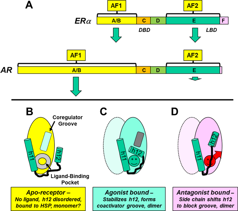
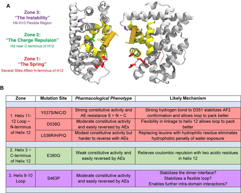
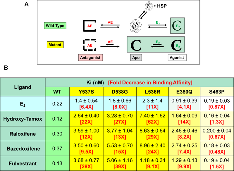
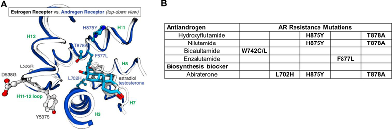
Oestrogen receptor-α (ERα), a key driver of breast cancer, normally requires oestrogen for activation. Mutations that constitutively activate ERα without the need for hormone binding are frequently found in endocrine-therapy-resistant breast cancer metastases and are associated with poor patient outcomes. The location of these mutations in the ER ligand-binding domain and their impact on receptor conformation suggest that they subvert distinct mechanisms that normally maintain the low basal state of wild-type ERα in the absence of hormone. Such mutations provide opportunities to probe fundamental issues underlying ligand-mediated control of ERα activity. Instructive contrasts between these ERα mutations and those that arise in the androgen receptor (AR) during anti-androgen treatment of prostate cancer highlight differences in how activation functions in ERs and AR control receptor activity, how hormonal pressures (deprivation versus antagonism) drive the selection of phenotypically different mutants, how altered protein conformations can reduce antagonist potency and how altered ligand-receptor contacts can invert the response that a receptor has to an agonist ligand versus an antagonist ligand. A deeper understanding of how ligand regulation of receptor conformation is linked to receptor function offers a conceptual framework for developing new anti-oestrogens that might be more effective in preventing and treating breast cancer.


Figures

Figure 1.
Figure 1.. Overview of nuclear receptor domain structure, activating functions, ligand-induced conformations, and dimer formation.
A. Domain structure of two nuclear hormone receptors, ERα and AR. The domains, shown to scale, are labeled A/B through F. The size of the green arrows associated with the activating functions (AF1 and AF2) illustrate their relative contribution to the transcriptional activity of ER or AR. The D domain is called the hinge, and the most C-terminal F domain is much more prominent in ERα than in AR. B-D. Schematic representation of three conformational states of the LBD of hormone-regulated nuclear receptors, highlighting the relative positions of the two carboxy-terminal helices, h11 and h12, and dimer states. B. In the unliganded or apo-receptor LBD, the LBP is empty (gray doughnut) and the coactivator groove (light blue rectangle) is incomplete and empty. C. With a bound agonist (green circle), h12 folds back, covering the LBP and generating the coactivator binding groove (dark gray rectangle). D. With a bound AE (red circle with arrow for a side chain), h12 moves to block the coactivator binding groove. The liganded LBDs (C and D) are represented as dimers; the second monomer is shown in lighter color, and conformational details are omitted. Although uncertain, the apo-LBD (B) is shown as a monomer.
Figure 2.
Figure 2.. Activating Mutations in the ERα LBD, Pharmacological Phenotypes, and Mechanisms.
A. Ribbon diagrams of two perspectives illustrating the three zones for the principal activating mutations in the ERα ligand-binding domain (LBD). The color codes (red, green, and purple) match the zone designations with the residues in each zone. All of these mutations alter residues that are far from the bound ligand, estradiol (ball and stick model). The region shown in yellow constitutes the coactivator binding groove and the orange cylinder, the coactivator LxxLL helical domain. (A movie showing a rotating 3D version of this structure is available – Supplemental Movie S1.) B. Tabular listing by zone of the principal activating mutations, with their pharmacological phenotype and likely functional mechanism. Constitutive activity and reversal here refers principally to the transcriptional activation functions of the receptor.
Figure 3.
Figure 3.. Schematic overview of estrogen agonist (E2) and antiestrogen (AE) binding to WT and mutant ERα LBDs and binding affinities of various antiestrogens to WT ERα and five mutant ERs.
A. The WT ERα LBD, which is unfolded in the absence of ligands and largely bound by heat shock proteins (HSP), gains extra energy upon binding of either E2 or AEs from formation of additional protein-protein contacts in the stably folded ligand complexes. Mutant ER LBDs are pre-folded in the agonist conformation, which reduces the binding affinity of both E2 and AEs, but AE binding is reduced to a greater extent because the agonist folding bias has to be overcome to access the antagonist conformation. Active species are highlighted in light green. B. Binding affinities for E2 and AEs to WT and constitutively active ERα mutants. The level of constitutive activity is indicated by the intensity of the shading (blue for WT ERα and yellow for the mutants). Estradiol binding is determined by a direct assay with [3H]estradiol by Scatchard analysis,. Antiestrogen binding was determined by a competitive binding assay using [3H]estradiol as a tracer and LBDs of WT and ERα mutants. Because the IC50 values from the competition assay are affected by the estradiol binding affinities, they have been converted to Ki values using the Cheng-Prusoff relationship. (Data on WT, Y537S and D538G ERα LBDs for estradiol, hydroxy-tamoxifen, and fulvestrant are updated values with more replicates from our published work,; the values on the other ERα mutants and for raloxifene and bazedoxifene were determined in our laboratories using the same published methodology,,.)
Figure 4.
Figure 4.. Comparative view of the locations of activating mutations in ER and in AR relative to the position of the ligand, and the relationship of AR mutations to specific AR antagonists.
A. A structural overlay of a portion of the LBDs of ER and estradiol (in gray) and testosterone (in blue). The sites of mutation in AR (702, 875, 877, and 878, blue residues) are within the ligand-binding pocket, close to the ligand, whereas the mutation sites in ER (536, 537, and 538, standard atom colored residues) are outside of the pocket, far from ligand contact. (Two rotating 3D movies are available: Supplemental Movie S2 shows that the resistance mutations in the AR LBD are within the LBP. Supplemental Movie S3 shows an overlap comparison of the two LBDs, contrasting the locations of the mutations relative to the ligand.) B. Specific AR mutations are associated with antiandrogens with different structures. (Because hydroxyflutamide and nilutamide have similar structures, they have similar sites of mutation.) While nominally a blocker of androgen biosynthesis from adrenal precursors, Abiraterone, and particularly its oxidized metabolite D4A, are also direct AR antagonist ligands., Abiraterone therapy also elevates levels of progestational ligands and suppresses corticosteroid production, necessitating corticosteroid supplementation. These three mutations reduce AR binding specificity and are activated by progestins and corticosteroids.

References

    1. Katzenellenbogen BS & Frasor J Therapeutic targeting in the estrogen receptor hormonal pathway. Semin. Oncol 31, 28–38, (2004). - PubMed
    1. Rugo HS et al. Endocrine Therapy for Hormone Receptor-Positive Metastatic Breast Cancer: American Society of Clinical Oncology Guideline. Journal of clinical oncology : official journal of the American Society of Clinical Oncology 34, 3069–3103, (2016). - PubMed
    1. Tryfonidis K, Zardavas D, Katzenellenbogen BS & Piccart M Endocrine treatment in breast cancer: Cure, resistance and beyond. Cancer treatment reviews 50, 68–81, (2016). - PubMed
    1. Smith DF & Toft DO Minireview: the intersection of steroid receptors with molecular chaperones: observations and questions. Mol. Endocrinol 22, 2229–2240, (2008). - PMC - PubMed
    1. Pratt WB & Toft DO Regulation of signaling protein function and trafficking by the hsp90/hsp70-based chaperone machinery. Exp Biol Med (Maywood) 228, 111–133, (2003). - PubMed

Publication types