Review
. 2018 Apr 14;24(14):1507-1520.
doi: 10.3748/wjg.v24.i14.1507.

Hepatitis B virus pre-S/S variants in liver diseases

Affiliations
Review

Hepatitis B virus pre-S/S variants in liver diseases

Bing-Fang Chen. World J Gastroenterol. .

Abstract

Chronic hepatitis B is a global health problem. The clinical outcomes of chronic hepatitis B infection include asymptomatic carrier state, chronic hepatitis (CH), liver cirrhosis (LC), and hepatocellular carcinoma (HCC). Because of the spontaneous error rate inherent to viral reverse transcriptase, the hepatitis B virus (HBV) genome evolves during the course of infection under the antiviral pressure of host immunity. The clinical significance of pre-S/S variants has become increasingly recognized in patients with chronic HBV infection. Pre-S/S variants are often identified in hepatitis B carriers with CH, LC, and HCC, which suggests that these naturally occurring pre-S/S variants may contribute to the development of progressive liver damage and hepatocarcinogenesis. This paper reviews the function of the pre-S/S region along with recent findings related to the role of pre-S/S variants in liver diseases. According to the mutation type, five pre-S/S variants have been identified: pre-S deletion, pre-S point mutation, pre-S1 splice variant, C-terminus S point mutation, and pre-S/S nonsense mutation. Their associations with HBV genotype and the possible pathogenesis of pre-S/S variants are discussed. Different pre-S/S variants cause liver diseases through different mechanisms. Most cause the intracellular retention of HBV envelope proteins and induction of endoplasmic reticulum stress, which results in liver diseases. Pre-S/S variants should be routinely determined in HBV carriers to help identify individuals who may be at a high risk of less favorable liver disease progression. Additional investigations are required to explore the molecular mechanisms of the pre-S/S variants involved in the pathogenesis of each stage of liver disease.

Keywords: Chronic hepatitis; Hepatitis B virus; Hepatocellular carcinoma; Liver cirrhosis; Pre-S deletion; Pre-S/S mutant; Splice variant; spPS1.


Conflict of interest statement

Conflict-of-interest statement: No potential conflicts of interest.

Figures
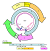
Figure 1
Figure 1
Genome structure and organization of hepatitis B virus. The relaxed-circular DNA genome of HBV with a complete minus strand and incomplete plus strand is shown (inner circle), along with the four main open reading frames (ORFs): pre-S/S; precore/core (pC/C); Pol, including four domains: TP, SP, reverse transcriptase (RT), and RNase H (RH); and X. The minus (-) and plus (+) DNA strands are marked. The HBV Pol and capped mRNA oligomer at the 5' end of the (-) and (+) strands as well as the DR-1 and DR-2 are illustrated. The space between the DR-1 and DR-2 is the "cohesive overlap region." The (+) strand is typically incomplete.
Figure 2
Figure 2
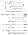
Gene expression of the pre-S/S gene in (A) wild-type hepatitis B virus and (B) pre-S/S variants: non-S promoter, S promoter, and spPS1.
Figure 3
Figure 3
Immune epitopes and functional domains within the hepatitis B virus pre-S region. The pre-S region consists of the pre-S1 and pre-S2 regions. The pre-S1 region contains 119 amino acids in HBV genotypes B or C and is further divided into two parts: the N half (amino acids 1-57) and C half (amino acids 58-119). The pre-S2 region contains 55 amino acids. The pre-S domain contains many B- or T-epitopes and exerts multiple functions, as illustrated. The N-half of pre-S1 contains a hepatocyte binding site essential for infection. The C-half of pre-S1 contains a heat-shock protein 70 (Hsc70) binding site and cytosolic anchorage determinant (CAD) vital for dual topology of L proteins as well as a nucleocapsid binding site (NBS) for virion morphogenesis. The C-half of pre-S1 also contains an S-promoter and CCAAT binding factor (CBF) binding site necessary for expression of the S gene. The pre-S2 region has a polymerized human serum albumin (pHSA) binging site and viral secretion (VS) site. Black triangle, myristylation at second amino acid; white triangle, N-link glycosylation at N-4 of the M protein; gray triangle, O-link glycosylation at T-37 of the M protein. B-epitopes: pS1-B1, pS1-B2, pS2-B1, pS2-B2, and pS2-B3. T-epitopes: pS1-T1 and pS2-T1. B- and T-epitope: pS1-BT.
Figure 4
Figure 4
Topology of wild-type small (S), medium (M), and large (L) envelope proteins. The predicted four membrane-spanning segments (indicated by rectangular boxes) of S project their N and C termini into the endoplasmic reticulum lumen (A). The M proteins exhibit a topology similar to the S protein with their N-terminal pre-S2 domain protruding into the endoplasmic reticulum (ER) lumen, whereas the L proteins display a dual topology. Upon cotranslational membrane integration, the pre-S domains of L proteins are initially located on the cytosolic side of the ER membrane (i-Pre-S); they are controlled by the dual topology site (indicated by an oval). During maturation (marked by the arrow), nearly half of mature L-protein molecules posttranslationally translocate their pre-S region to the luminal space (e-Pre-S). The nucleocapsid (N) binding sites in the pre-S and S region are indicated by the white curved box. (B) The L-protein topology of pre-S/S variants. The nucleocapsid variant demonstrates a dual topology, and the topology variants and spPS1 variants display a uniform topology. The broken line indicates deletion, and “a” indicates “a” determinant.
Figure 5
Figure 5
Proposed model for the generation of pre-S/S variants and their possible roles in liver damage and carcinogenesis. HBV: Hepatitis B virus; ER: Endoplasmic reticulum; NBS: Nucleocapsid binding site; VS: Viral secretion.

References

    1. Seeger C, Mason WS. Hepatitis B virus biology. Microbiol Mol Biol Rev. 2000;64:51–68. - PMC - PubMed
    1. Kao JH, Chen DS. Global control of hepatitis B virus infection. Lancet Infect Dis. 2002;2:395–403. - PubMed
    1. Liu CJ, Kao JH. Global perspective on the natural history of chronic hepatitis B: role of hepatitis B virus genotypes A to J. Semin Liver Dis. 2013;33:97–102. - PubMed
    1. Lin CL, Kao JH. The clinical implications of hepatitis B virus genotype: Recent advances. J Gastroenterol Hepatol. 2011;26 Suppl 1:123–130. - PubMed
    1. Sugauchi F, Ohno T, Orito E, Sakugawa H, Ichida T, Komatsu M, Kuramitsu T, Ueda R, Miyakawa Y, Mizokami M. Influence of hepatitis B virus genotypes on the development of preS deletions and advanced liver disease. J Med Virol. 2003;70:537–544. - PubMed

MeSH terms

Substances