. 2018 Mar 12:9:473.
doi: 10.3389/fimmu.2018.00473. eCollection 2018.

Long Non-Coding RNA HOXA Transcript Antisense RNA Myeloid-Specific 1-HOXA1 Axis Downregulates the Immunosuppressive Activity of Myeloid-Derived Suppressor Cells in Lung Cancer

Affiliations

Long Non-Coding RNA HOXA Transcript Antisense RNA Myeloid-Specific 1-HOXA1 Axis Downregulates the Immunosuppressive Activity of Myeloid-Derived Suppressor Cells in Lung Cancer

Xinyu Tian et al. Front Immunol. .

Erratum in

Abstract

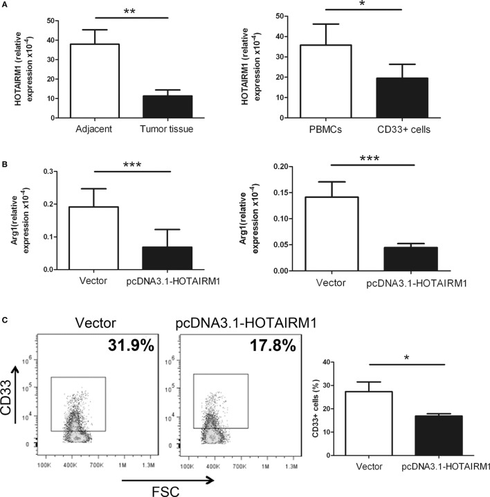
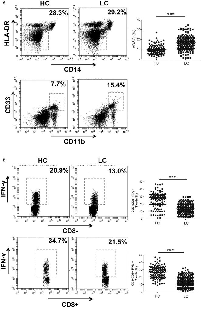
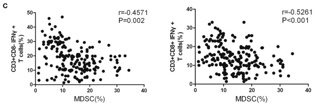
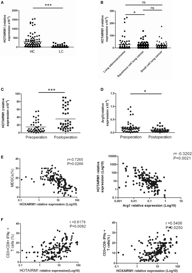
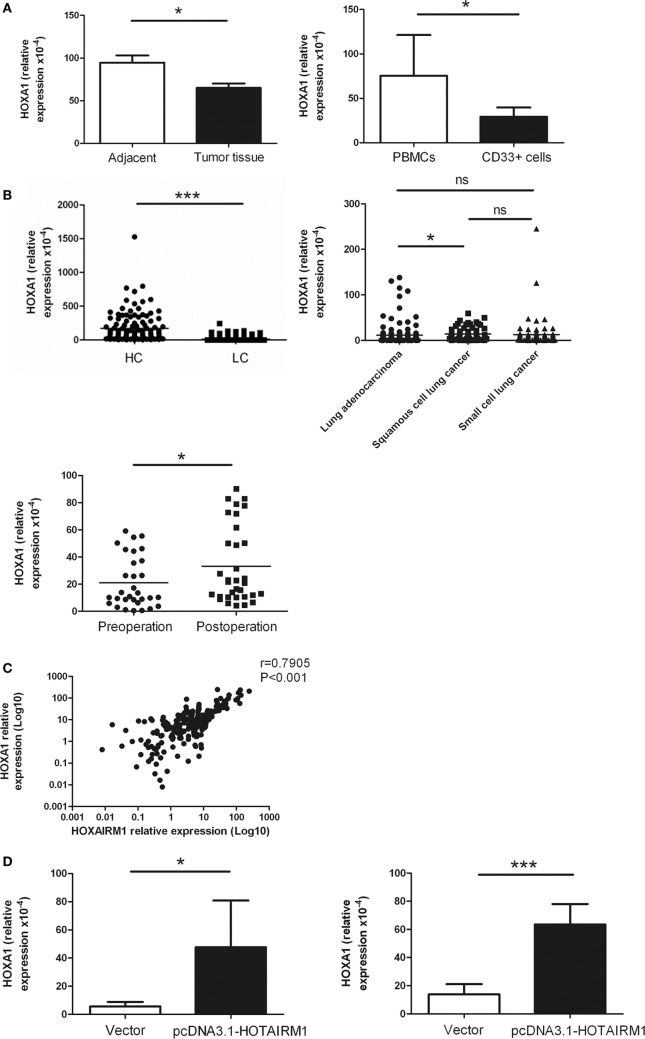
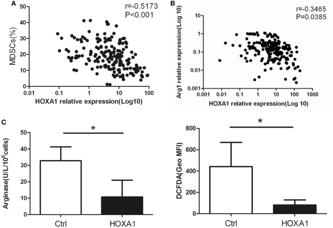
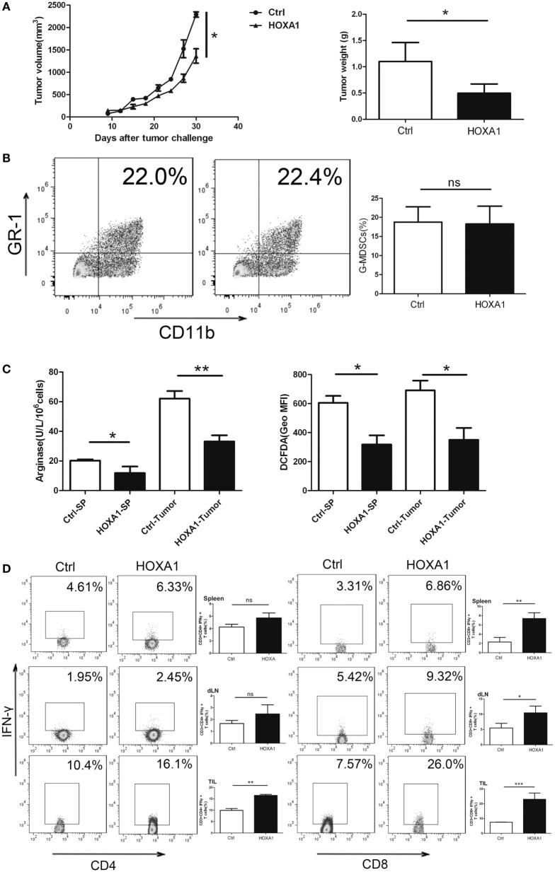
HOXA transcript antisense RNA myeloid-specific 1 (HOTAIRM1) is a long non-coding RNA that has been shown to be a key regulator of myeloid cell development by targeting HOXA1. Myeloid-derived suppressor cells (MDSCs) are a heterogeneous population of immature myeloid cells that possess immunosuppressive function. However, the impact of HOTAIRM1 on the development of MDSCs remains unknown. In this study, we demonstrated that HOTAIRM1 was expressed in MDSCs and that overexpression of HOTAIRM1 could downregulate the expression of suppressive molecules in MDSCs. In addition, HOTAIRM1 levels were observed to be decreased in the peripheral blood cells of lung cancer patients compared with those of healthy controls. By analyzing HOTAIRM1 expression levels in different types of lung cancer, we found that HOTAIRM1 was mainly expressed in lung adenocarcinoma. Finally, it was confirmed that HOTAIRM1 could enhance the expression of HOXA1 in MDSCs and that high levels of HOXA1, the target gene of HOTAIRM1, could delay tumor progression and enhance the antitumor immune response by downregulating the immunosuppression of MDSCs. Taken together, this study illustrates that HOTAIRM1/HOXA1 downregulates the immunosuppressive function of MDSCs and may be a potential therapeutic target in lung cancer.

Keywords: HOXA transcript antisense RNA myeloid-specific 1; HOXA1; long non-coding RNA; lung cancer; myeloid-derived suppressor cells.


Figures

Figure 1
Figure 1
HOXA transcript antisense RNA myeloid-specific 1 (HOTAIRM1) can inhibit myeloid-derived suppressor cell (MDSC) development in lung cancer. (A) The expression level of HOTAIRM1 in MDSCs isolated from tumor tissues of patients with lung cancer (left) or induced from peripheral blood mononuclear cells (PBMCs) of healthy donors with GM-CSF (40 ng/mL) + IL-1β (40 ng/mL) (right) was detected with qRT-PCR. (B) The MDSCs isolated from tumor tissues of patients with lung cancer (left) or induced from PBMCs of healthy donors with GM-CSF (40 ng/mL) + IL-1β (40 ng/mL) (right) were transfected with pcDNA3.1-HOTAIRM1, and arginase 1 (Arg1) levels were detected by qRT-PCR. (C) The effect of HOTAIRM1 overexpression on the induction of MDSCs (***P < 0.001, **P < 0.01, and *P < 0.05).
Figure 2
Figure 2
The proportion of myeloid-derived suppressor cells (MDSCs) is negatively correlated with the percentage of Th1/CTL cells in the peripheral blood of lung cancer patients. (A) The proportion of CD11b+CD33+HLA-DRCD14 MDSCs in the peripheral blood of healthy controls (n = 300) and lung cancer patients (n = 285). (B) The proportions of CD3+CD8-IFN-γ+ Th1 and CD3+CD8+IFN-γ+ CTL cells in the peripheral blood of healthy controls (n = 285) and patients with lung cancer (n = 300). (C) The correlation between the proportion of MDSCs and the percentage of CD3+CD8-IFN-γ+ Th1/CD3+CD8+IFN-γ+ CTL cells in the peripheral blood of patients with lung cancer (n = 300) (***P < 0.001).
Figure 2
Figure 2
The proportion of myeloid-derived suppressor cells (MDSCs) is negatively correlated with the percentage of Th1/CTL cells in the peripheral blood of lung cancer patients. (A) The proportion of CD11b+CD33+HLA-DRCD14 MDSCs in the peripheral blood of healthy controls (n = 300) and lung cancer patients (n = 285). (B) The proportions of CD3+CD8-IFN-γ+ Th1 and CD3+CD8+IFN-γ+ CTL cells in the peripheral blood of healthy controls (n = 285) and patients with lung cancer (n = 300). (C) The correlation between the proportion of MDSCs and the percentage of CD3+CD8-IFN-γ+ Th1/CD3+CD8+IFN-γ+ CTL cells in the peripheral blood of patients with lung cancer (n = 300) (***P < 0.001).
Figure 3
Figure 3
The expression of HOXA transcript antisense RNA myeloid-specific 1 (HOTAIRM1) was significantly decreased in the peripheral blood of patients with lung cancer. (A) The expression of HOTAIRM1 in the peripheral blood of lung cancer patients (n = 300). (B) The expression of HOTAIRM1 in the peripheral blood of patients with different types of lung cancer (n = 300). (C) HOTAIRM1 expression levels in the peripheral blood of lung cancer patients pre- and post-operation (n = 42). (D) Arginase 1 (Arg1) levels in the peripheral blood of lung cancer patients pre- and postoperation (n = 42). (E) Correlation analysis between HOTAIRM1 levels and the proportion of myeloid-derived suppressor cells (MDSCs) or the Arg1 levels in the peripheral blood of patients with lung cancer (n = 300). (F) The correlation between the expression of HOTAIRM1 and the proportion of CD3+CD8-IFN-γ+/CD3+CD8+IFN-γ+ cells in the peripheral blood of lung cancer patients (n = 300) (***P < 0.001 and *P < 0.05).
Figure 4
Figure 4
HOXA transcript antisense RNA myeloid-specific 1 (HOTAIRM1) enhances the expression of HOXA1. (A) The expression of HOXA1 in myeloid-derived suppressor cells (MDSCs) isolated from tumor tissues of patients with lung cancer (left) or induced from peripheral blood mononuclear cells (PBMCs) of healthy donors with GM-CSF (40 ng/mL) + IL-1β (40 ng/mL) (right) was detected by qRT-PCR. (B) HOXA1 levels in the peripheral blood of healthy controls (n = 285) and patients with lung cancer (n = 300), and the expression of HOXA1 in the peripheral blood of different types of lung cancer (n = 300). (C) HOXA1 levels in the peripheral blood of lung cancer patients pre- and postoperation (n = 42). (D) The MDSCs isolated from tumor tissues of patients with lung cancer (left) or induced from PBMCs of healthy donors with GM-CSF (40 ng/mL) + IL-1β (40 ng/mL) (right) were transfected with pcDNA3.1-HOTAIRM1, and HOXA1 levels were detected by qRT-PCR (***P < 0.001, *P < 0.05, and ns, no significance).
Figure 5
Figure 5
HOXA1 is associated with the immunosuppression of myeloid-derived suppressor cells (MDSCs). (A) Correlation analysis between HOXA1 levels and the proportion of MDSCs in the peripheral blood of lung cancer patients (n = 300). (B) Correlation analysis between HOXA1 levels and arginase 1 (Arg1) levels in the peripheral blood of lung cancer patients (n = 300). (C) Arg1 activity and reactive oxygen species production in MDSCs from the spleen of tumor-bearing mice following transfection with pcDNA3.1-HOXA1 (*P < 0.05).
Figure 6
Figure 6
Overexpression of HOXA1 can delay tumor progression and enhance the antitumor immune response in tumor-bearing mice. Groups of mice bearing established LLC cells were intratumorally injected with pcDNA3.1-HOXA1 every 3 days for 3 weeks. (A) Tumor volume and weight were measured at the indicated times. (B) The proportion of CD11b+Gr1+ MDSCs in tumor tissue was analyzed by flow cytometry. (C) Arginase 1 activity and reactive oxygen species production in MDSCs sorted from the spleen and tumor tissues were detected. (D) The proportions of CD3+CD4+IFN-γ+ Th1 and CD3++CD8+IFN-γ+ CTL cells from spleens, draining lymph nodes and tumor tissue were analyzed by flow cytometry (***P < 0.001, **P < 0.01, *P < 0.05, and ns, no significance).

References

    1. Aerts JG, Hegmans JP. Tumor-specific cytotoxic T cells are crucial for efficacy of immunomodulatory antibodies in patients with lung cancer. Cancer Res (2013) 73(8):2381–8.10.1158/0008-5472.CAN-12-3932 - DOI - PubMed
    1. Blandin Knight S, Crosbie PA, Balata H, Chudziak J, Hussell T, Dive C. Progress and prospects of early detection in lung cancer. Open biology (2017) 7(9):170070.10.1098/rsob.170070 - DOI - PMC - PubMed
    1. Cai Q, Luo HL, Gao XC, Xiong CJ, Tong F, Zhang RG, et al. Clinical features and prognostic factors of small cell lung cancer: a retrospective study in 148 patients. J Huazhong Univ Sci Technolog Med Sci (2016) 36(6):916–22.10.1007/s11596-016-1684-6 - DOI - PubMed
    1. Kang M, Li Y, Zhao Y, He S, Shi J. miR-33a inhibits cell proliferation and invasion by targeting CAND1 in lung cancer. Clin Transl Oncol (2017).10.1007/s12094-017-1730-2 - DOI - PubMed
    1. Chalela R, Curull V, Enriquez C, Pijuan L, Bellosillo B, Gea J. Lung adenocarcinoma: from molecular basis to genome-guided therapy and immunotherapy. J Thorac Dis (2017) 9(7):2142–58.10.21037/jtd.2017.06.20 - DOI - PMC - PubMed

Publication types

MeSH terms