. 2018 Mar 23;9(22):15942-15951.
doi: 10.18632/oncotarget.24639.

Ectopic expression of transcription factor BATF3 induces B-cell lymphomas in a murine B-cell transplantation model

Affiliations

Ectopic expression of transcription factor BATF3 induces B-cell lymphomas in a murine B-cell transplantation model

Christian Weiser et al. Oncotarget. .

Abstract

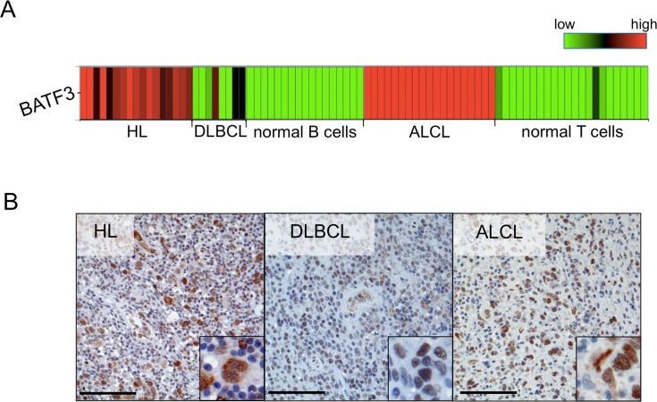
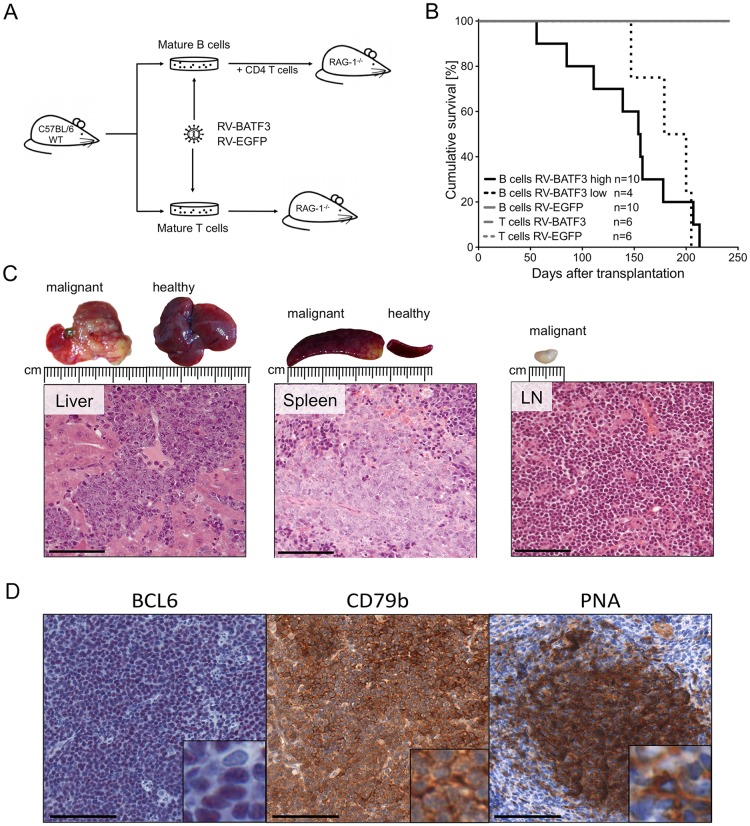
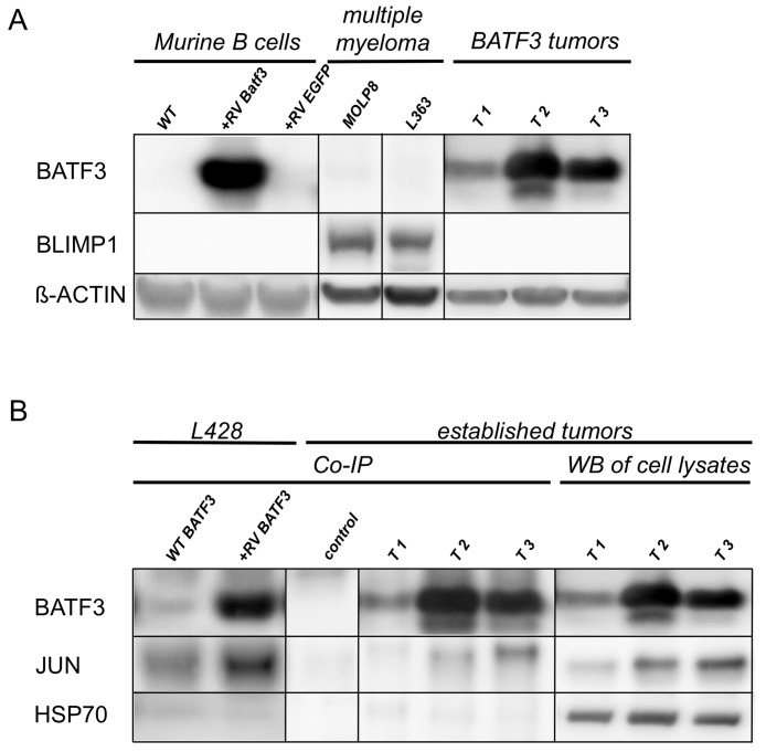
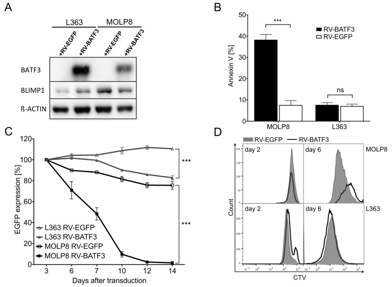
The mechanisms involved in malignant transformation of mature B and T lymphocytes are still poorly understood. In a previous study, we compared gene expression profiles of the tumor cells of Hodgkin lymphoma (HL) and anaplastic large cell lymphoma (ALCL) to their normal cellular counterparts and found the basic leucine zipper protein ATF-like 3 (BATF3) to be significantly upregulated in the tumor cells of both entities. To assess the oncogenic potential of BATF3 in lymphomagenesis and to dissect the molecular interactions of BATF3 in lymphoma cells, we retrovirally transduced murine mature T and B cells with a BATF3-encoding viral vector and transplanted each population into Rag1-deficient recipients. Intriguingly, BATF3-expressing B lymphocytes readily induced B-cell lymphomas after characteristic latencies, whereas T-cell transplanted animals remained healthy throughout the observation time. Further analyses revealed a germinal center B-cell-like phenotype of most BATF3-initiated lymphomas. In a multiple myeloma cell line, BATF3 inhibited BLIMP1 expression, potentially illuminating an oncogenic action of BATF3 in B-cell lymphomagenesis. In conclusion, BATF3 overexpression induces malignant transformation of mature B cells and might serve as a potential target in B-cell lymphoma treatment.

Keywords: B-cell immunology; B-cell lymphoma; BATF3; germinal center; oncogene.


Conflict of interest statement

CONFLICTS OF INTEREST The authors declare no competing financial interests.

Figures

Figure 1
Figure 1. BATF3 is aberrantly expressed in several lymphoma entities
(A) Heatmap highlighting the upregulated gene expression of BATF3 in Hodgkin lymphoma (HL), anaplastic large cell lymphoma (ALCL) and some diffuse large B-cell lymphoma (DLBCL) cases, when compared to the expression in normal human B (mature, germinal center and plasma cells) and T cells (mature CD4+ and CD8+ cells). BATF3 gene expression data were extracted from Brune et al. [16] and Eckerle et al. [15]. (B) Representative immunohistochemistry of BATF3 in HL, DLBCL and ALCL, displaying nuclear BATF3 expression. Bars 100 μm (200 x, insert 400 x).
Figure 2
Figure 2. Overexpression of BATF3 in mature B cells results in lymphomagenesis
(A) Experimental design and transplantation model. Wild type (WT) C57BL/6 mice were used as donors for mature B and T cells. Isolated cells were stimulated in cell culture, retrovirally transduced and transplanted into Rag1-deficient recipient mice. B-cells were co-transplanted with freshly isolated, TCR quasi-monoclonal, CD4+ T cells from OT-II mice. Mice were monitored by blood sampling and monitored for lymphoma development. (B) Survival of T- and B-cell transplanted recipients. Cohorts transplanted with BATF3-expressing B cells (black solid and dotted lines) developed lymphomas after different latencies. Animals that received highly BATF3-transduced B cells (black solid line) succumbed earlier to lymphoma than recipients of low BATF3-expressing B cells (black dotted line). One of the mice transplanted with cells transduced with high levels of BATF3 vector died of lymphoma, but could not be included in the detailed characterization of the lymphomas because of insufficient quality of the necrotic tissue. Recipients of BATF3-expressing T cells and EGFP-control cells did not show any signs of malignancy during the observation period (gray lines). (C) Representative histologies of a BATF3-induced B-cell lymphoma. Necropsy of diseased animals revealed massive infiltration of tumor cells into the liver, spleen and lymph nodes. Hematoxilin and eosin stained tumors demonstrated blastoid infiltrates and a destruction of the normal, organ-specific architecture. Bars represent 100 μm. (D) Representative immunohistochemical stainings of BATF3-induced tumors. Sections of tumor-infiltrated spleen of BATF3-induced lymphomas were stained for the B-cell marker CD79b and the GC B-cell markers BCL6 and PNA. Bars represent 100 μm.
Figure 3
Figure 3. BATF3 interacts with JUN in BLIMP1-negative, BATF3-induced tumor cell lines
(A) Western blot analysis of LPS-stimulated, freshly transduced murine B-cells and established murine cell lines from BATF3-induced tumors (lanes T1-3) for the expression of BATF3, and BLIMP1. RV indicates retroviral transduction with BATF3 or EGFP as a control. Multiple myeloma cell lines MOLP8 and L363 were used as positive control for BLIMP1 expression. (B) Co-IP of BATF3 and JUN with HL cell line L428 and BATF3-induced murine B-cell tumor cell lines. L428 cells showed an endogenous expression of BATF3, therefore JUN was immunoprecipitated in wild type (WT) L428 cells (WT BATF3 lane). As expected, retroviral expression of BATF3 in L428 cells even increased the amount of retained JUN protein after Co-IP (+RV-BATF3 lane). Most interestingly, interaction of BATF3 and JUN was demonstrated in BATF3-induced tumors (lanes T1-3). Western blot of the corresponding whole lysates and after Co-IP without specific antibodies served as controls. HSP70 was used as negative control for Co-IP experiments. All Co-IP experiments were performed with a BATF3-specific antibody.
Figure 4
Figure 4. BATF3-overexpression leads to downregulation of BLIMP1 and apoptosis in multiple myeloma (MM) cell line MOLP8
(A) Expression of BLIMP1 and BATF3 in MM cell lines MOLP8 and L363 after retroviral transduction with BATF3 (+RV-BATF3) or EGFP (+RV-EGFP) as a control. Ectopic BATF3-expression induced a downregulation of BLIMP1 in MOLP8 cells, whereas BLIMP1 was found to be upregulated in L363 cells after BATF3-transduction. (B) BATF3-induced apoptotic cell death in MM cell line MOLP8. Seven days after transduction with BATF3, MOLP8 cells showed an increased fraction of apoptotic cells (left black bar) when compared to control-gene transduced cells (left open bar). BATF3-overexpression in L363 cells did not lead to increased apoptosis (right black and open bars). (C) BATF3-expression dramatically impaired cell growth of MM cell line MOLP8 (black line with solid square). On the other hand, control-gene modified MOLP8 cells (black line with open square) and transduced L363 cells (grey line with solid or open triangles) did not show reduced expansion. (D) Proliferation of MM cell lines MOLP8 and L363 after transduction with BATF3 or control gene EGFP. Directly after transduction, both cell lines were labeled with the cell-division tracker CellTrace Violet (CTV) and were analyzed by flow cytometry on days 2 and 7. Subsequently, flow cytometric analysis on day 7 revealed that BATF3-expressing MOLP8 cells (solid line) compared to EGFP-modified control cells (grey area) showed a reduced proliferation. L363 cells were not affected in their proliferation capacity after BATF3-overexpression. Error bars represent standard deviation. Statistical significance was established with a paired t test. All experiments were performed in triplicates. ***, P < 0.0001, ns, not significant

References

    1. Küppers R. Mechanisms of B-cell lymphoma pathogenesis. Nat Rev Cancer. 2005;5:251–262. - PubMed
    1. Küppers R, Dalla-Favera R. Mechanisms of chromosomal translocations in B cell lymphomas. Oncogene. 2001;20:5580–5594. - PubMed
    1. MacLennan IC. Germinal centers. Annu Rev Immunol. 1994;12:117–139. - PubMed
    1. Seifert M, Scholtysik R, Küppers R. Origin and pathogenesis of B cell lymphomas. Methods Mol Biol. 2013;971:1–25. - PubMed
    1. Shaffer AL, Lin KI, Kuo TC, Yu X, Hurt EM, Rosenwald A, Giltnane JM, Yang L, Zhao H, Calame K, Staudt LM. Blimp-1 orchestrates plasma cell differentiation by extinguishing the mature B cell gene expression program. Immunity. 2002;17:51–62. - PubMed