

Isolation of Extracellular Vesicles: General Methodologies and Latest Trends
- PMID: 29662902
- PMCID: PMC5831698
- DOI: 10.1155/2018/8545347
Isolation of Extracellular Vesicles: General Methodologies and Latest Trends
Abstract
Background: Extracellular vesicles (EVs) play an essential role in the communication between cells and transport of diagnostically significant molecules. A wide diversity of approaches utilizing different biochemical properties of EVs and a lack of accepted protocols make data interpretation very challenging.
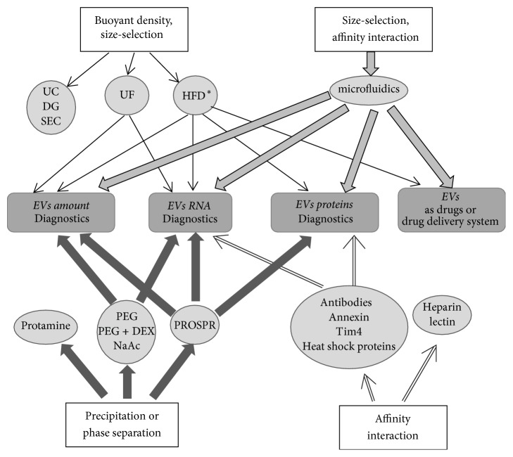
Scope of review: This review consolidates the data on the classical and state-of-the-art methods for isolation of EVs, including exosomes, highlighting the advantages and disadvantages of each method. Various characteristics of individual methods, including isolation efficiency, EV yield, properties of isolated EVs, and labor consumption are compared.
Major conclusions: A mixed population of vesicles is obtained in most studies of EVs for all used isolation methods. The properties of an analyzed sample should be taken into account when planning an experiment aimed at studying and using these vesicles. The problem of adequate EVs isolation methods still remains; it might not be possible to develop a universal EV isolation method but the available protocols can be used towards solving particular types of problems.
General significance: With the wide use of EVs for diagnosis and therapy of various diseases the evaluation of existing methods for EV isolation is one of the key problems in modern biology and medicine.
Figures
References
-
- Tamkovich S. N., Tutanov O. S., Laktionov P. P. Exosomes: Generation, structure, transport, biological activity, and diagnostic application. Biologicheskie membrany. 2016;33(3):163–175. doi: 10.7868/S0233475516020122. (Rus). - DOI
Publication types
MeSH terms
LinkOut - more resources
Full Text Sources
Other Literature Sources
Research Materials
