

Molecular interactions at the surface of extracellular vesicles
- PMID: 29663027
- PMCID: PMC6208672
- DOI: 10.1007/s00281-018-0682-0
Molecular interactions at the surface of extracellular vesicles
Abstract
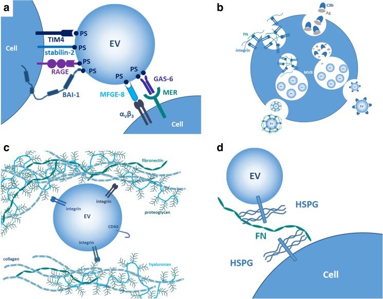
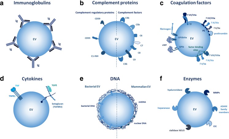
Extracellular vesicles such as exosomes, microvesicles, apoptotic bodies, and large oncosomes have been shown to participate in a wide variety of biological processes and are currently under intense investigation in many different fields of biomedicine. One of the key features of extracellular vesicles is that they have relatively large surface compared to their volume. Some extracellular vesicle surface molecules are shared with those of the plasma membrane of the releasing cell, while other molecules are characteristic for extracellular vesicular surfaces. Besides proteins, lipids, glycans, and nucleic acids are also players of extracellular vesicle surface interactions. Being secreted and present in high number in biological samples, collectively extracellular vesicles represent a uniquely large interactive surface area which can establish contacts both with cells and with molecules in the extracellular microenvironment. Here, we provide a brief overview of known components of the extracellular vesicle surface interactome and highlight some already established roles of the extracellular vesicle surface interactions in different biological processes in health and disease.
Keywords: Drug delivery; Extracellular matrix; Extracellular vesicle; Interactome; Surface.
Figures
References
-
- Yanez-Mo M, Siljander PR, Andreu Z, Zavec AB, Borras FE, Buzas EI, Buzas K, Casal E, Cappello F, Carvalho J, Colas E, Cordeiro-da Silva A, Fais S, Falcon-Perez JM, Ghobrial IM, Giebel B, Gimona M, Graner M, Gursel I, Gursel M, Heegaard NH, Hendrix A, Kierulf P, Kokubun K, Kosanovic M, Kralj-Iglic V, Kramer-Albers EM, Laitinen S, Lasser C, Lener T, Ligeti E, Line A, Lipps G, Llorente A, Lotvall J, Mancek-Keber M, Marcilla A, Mittelbrunn M, Nazarenko I, Nolte-'t Hoen EN, Nyman TA, O'Driscoll L, Olivan M, Oliveira C, Pallinger E, Del Portillo HA, Reventos J, Rigau M, Rohde E, Sammar M, Sanchez-Madrid F, Santarem N, Schallmoser K, Ostenfeld MS, Stoorvogel W, Stukelj R, Van der Grein SG, Vasconcelos MH, Wauben MH, De Wever O. Biological properties of extracellular vesicles and their physiological functions. J Extracell Vesicles. 2015;4:27066. - PMC - PubMed
-
- Di Vizio D, Morello M, Dudley AC, Schow PW, Adam RM, Morley S, Mulholland D, Rotinen M, Hager MH, Insabato L, Moses MA, Demichelis F, Lisanti MP, Wu H, Klagsbrun M, Bhowmick NA, Rubin MA, D'Souza-Schorey C, Freeman MR. Large oncosomes in human prostate cancer tissues and in the circulation of mice with metastatic disease. Am J Pathol. 2012;181:1573–1584. - PMC - PubMed
-
- Lotvall J, Hill AF, Hochberg F, Buzas EI, Di Vizio D, Gardiner C, Gho YS, Kurochkin IV, Mathivanan S, Quesenberry P, Sahoo S, Tahara H, Wauben MH, Witwer KW, Thery C. Minimal experimental requirements for definition of extracellular vesicles and their functions: a position statement from the International Society for Extracellular Vesicles. J Extracell Vesicles. 2014;3:26913. - PMC - PubMed
Publication types
MeSH terms
Grants and funding
LinkOut - more resources
Full Text Sources
Other Literature Sources
