

GFAP-Positive Progenitor Cell Production is Concentrated in Specific Encephalic Regions in Young Adult Mice
- PMID: 29663175
- PMCID: PMC6129256
- DOI: 10.1007/s12264-018-0228-4
GFAP-Positive Progenitor Cell Production is Concentrated in Specific Encephalic Regions in Young Adult Mice
Abstract
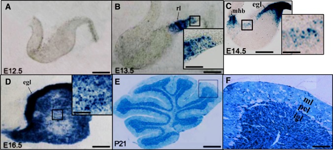
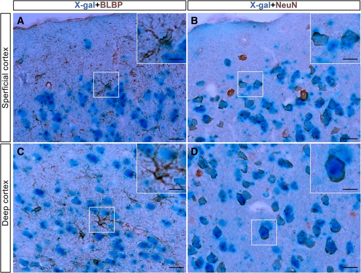
Previous genetic fate-mapping studies have indicated that embryonic glial fibrillary acidic protein-positive (GFAP+) cells are multifunctional progenitor/neural stem cells that can produce astrocytes as well as neurons and oligodendrocytes throughout the adult mouse central nervous system (CNS). However, emerging evidence from recent studies indicates that GFAP+ cells adopt different cell fates and generate different cell types in different regions. Moreover, the fate of GFAP+ cells in the young adult mouse CNS is not well understood. In the present study, hGFAP-Cre/R26R transgenic mice were used to investigate the lineage of embryonic GFAP+ cells in the young adult mouse CNS. At postnatal day 21, we found that GFAP+ cells mainly generated NeuN+ neurons in the cerebral cortex (both ventral and dorsal), hippocampus, and cerebellum. Strangely, these cells were negative for the Purkinje cell marker calbindin in the cerebellum and the neuronal marker NeuN in the thalamus. Thus, contrary to previous studies, our genetic fate-mapping revealed that the cell fate of embryonic GFAP+ cells at the young adult stage is significantly different from that at the adult stage.
Keywords: Astrocytes; Cell fate; GFAP; Neural stem cells; Neurons.
Conflict of interest statement
All authors claim that there are no conflicts of interest.
Figures


Similar articles
-
Early postnatal GFAP-expressing cells produce multilineage progeny in cerebrum and astrocytes in cerebellum of adult mice.Brain Res. 2013 Sep 26;1532:14-20. doi: 10.1016/j.brainres.2013.08.003. Epub 2013 Aug 9. Brain Res. 2013. PMID: 23939222
-
Early postnatal astroglial cells produce multilineage precursors and neural stem cells in vivo.J Neurosci. 2006 Aug 16;26(33):8609-21. doi: 10.1523/JNEUROSCI.2532-06.2006. J Neurosci. 2006. PMID: 16914687 Free PMC article.
-
Neonatal SVZ EGFP-labeled cells produce neurons in the olfactory bulb and astrocytes in the cerebral cortex by in-vivo electroporation.Neuroreport. 2013 May 8;24(7):381-7. doi: 10.1097/WNR.0b013e328360f7ef. Neuroreport. 2013. PMID: 23568218
-
GFAP-positive progenitor cells produce neurons and oligodendrocytes throughout the CNS.Mol Cell Neurosci. 2006 Apr;31(4):676-84. doi: 10.1016/j.mcn.2005.12.006. Epub 2006 Feb 3. Mol Cell Neurosci. 2006. PMID: 16458536
-
AQP4 gene deletion in mice does not alter blood-brain barrier integrity or brain morphology.Neuroscience. 2009 Jul 7;161(3):764-72. doi: 10.1016/j.neuroscience.2009.03.069. Epub 2009 Apr 5. Neuroscience. 2009. PMID: 19345723 Review.
Cited by
-
Effects of C5a and Receptor CD88 on Glutamate and N-Methyl-D-Aspartic Acid Receptor Expression in the Mouse Model of Optic Neuromyelitis.Comput Math Methods Med. 2022 Apr 25;2022:4997393. doi: 10.1155/2022/4997393. eCollection 2022. Comput Math Methods Med. 2022. PMID: 35509858 Free PMC article.
-
Expression Patterns of Inducible Cre Recombinase Driven by Differential Astrocyte-Specific Promoters in Transgenic Mouse Lines.Neurosci Bull. 2020 May;36(5):530-544. doi: 10.1007/s12264-019-00451-z. Epub 2019 Dec 11. Neurosci Bull. 2020. PMID: 31828740 Free PMC article.
-
Regulation of embryonic and adult neurogenesis by Ars2.Development. 2020 Jan 22;147(2):dev180018. doi: 10.1242/dev.180018. Development. 2020. PMID: 31969356 Free PMC article.
-
Tooth-Cutting-Induced Maxillary Malocclusion Exacerbates Cognitive Deficit in a Mouse Model of Vascular Dementia.Brain Sci. 2023 May 10;13(5):781. doi: 10.3390/brainsci13050781. Brain Sci. 2023. PMID: 37239252 Free PMC article.
-
Neuroprotective effects of methanolic extract from Chuanxiong Rhizoma in mice with middle cerebral artery occlusion-induced ischemic stroke: suppression of astrocyte- and microglia-related inflammatory response.BMC Complement Med Ther. 2024 Apr 4;24(1):140. doi: 10.1186/s12906-024-04454-w. BMC Complement Med Ther. 2024. PMID: 38575941 Free PMC article.
References
-
- Malatesta P, Harfuss E, Gotz M. Isolation of radial glial cells by fluorescent-activated cell sorting reveals a neuronal lineage. Development. 2000;127:5253–5263. - PubMed
MeSH terms
Substances
LinkOut - more resources
Full Text Sources
Other Literature Sources
Miscellaneous
