

Interactions of β tubulin isotypes with glutathione in differentiated neuroblastoma cells subject to oxidative stress
- PMID: 29663696
- PMCID: PMC6191380
- DOI: 10.1002/cm.21447
Interactions of β tubulin isotypes with glutathione in differentiated neuroblastoma cells subject to oxidative stress
Abstract
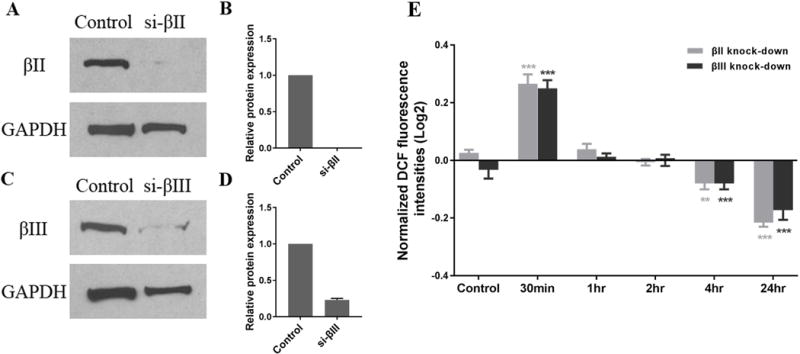
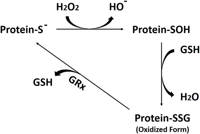
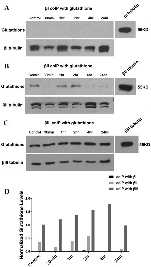
Microtubules are a major component of the neuronal cytoskeleton. Tubulin, the subunit protein of microtubules, is an α/β heterodimer. Both α and β exist as families of isotypes, whose members are encoded by different genes and have different amino acid sequences. The βII and βIII isotypes are very prominent in the nervous system. Our previous work has suggested that βII may play a role in neuronal differentiation, but the role of βIII in neurons is not well understood. In the work reported here, we examined the roles of the different β-tubulin isotypes in response to glutamate/glycine treatment, and found that both βII and βIII bind to glutathione in the presence of ROS, especially βIII. In contrast, βI did not bind to glutathione. Our results suggest that βII and βIII, but especially βIII, may play an important role in the response of neuronal cells to stress. In view of the high levels of βII and βIII expressed in the nervous system it is conceivable that these tubulin isotypes may use their sulfhydryl groups to scavenge ROS and protect neuronal cells against oxidative stress.
Keywords: glutathionylation; microtubules; oxidative stress; tubulin.
© 2018 Wiley Periodicals, Inc.
Conflict of interest statement
Conflict of Interest: Dr. Ludueña owns 500,000 shares in the company Oncovista Innovative Therapies, Inc.
Figures
References
-
- Bai RL, Lin CM, Nguyen NY, Liu TY, Hamel E. Identification of the cysteine residue of beta-tubulin alkylated by the antimitotic agent 2,4-dichlorobenzyl thiocyanate, facilitated by separation of the protein subunits of tubulin by hydrophobic column chromatography. Biochemistry. 1989;28:5606–5612. - PubMed
-
- Banan A, Fields JZ, Decker H, Zhang Y, Keshavarzian A. Nitric oxide and its metabolites mediate ethanol-induced microtubule disruption and intestinal barrier dysfunction. J Pharmacol Exp Ther. 2000;294:997–1008. - PubMed
-
- Banerjee A, Jordan MA, Luduena RF. Interaction of reduced glutathione with bovine brain tubulin. Biochem Biophys Res Commun. 1985;128:506–512. - PubMed
-
- Banerjee A, Roach MC, Trcka P, Luduena RF. Increased microtubule assembly in bovine brain tubulin lacking the type III isotype of beta-tubulin. J Biol Chem. 1990;265:1794–1799. - PubMed
-
- Banerjee A, Roach MC, Trcka P, Luduena RF. Preparation of a monoclonal antibody specific for the class IV isotype of beta-tubulin. Purification and assembly of alpha beta II, alpha beta III, and alpha beta IV tubulin dimers from bovine brain. J Biol Chem. 1992;267:5625–5630. - PubMed
Publication types
MeSH terms
Substances
Grants and funding
LinkOut - more resources
Full Text Sources
Other Literature Sources
Medical
