. 2018 Jun;7(11):e1701347.
doi: 10.1002/adhm.201701347. Epub 2018 Apr 16.

Patient-Specific Bioinks for 3D Bioprinting of Tissue Engineering Scaffolds

Affiliations

Patient-Specific Bioinks for 3D Bioprinting of Tissue Engineering Scaffolds

Negar Faramarzi et al. Adv Healthc Mater. 2018 Jun.

Abstract

Bioprinting has emerged as a promising tool in tissue engineering and regenerative medicine. Various 3D printing strategies have been developed to enable bioprinting of various biopolymers and hydrogels. However, the incorporation of biological factors has not been well explored. As the importance of personalized medicine is becoming more clear, the need for the development of bioinks containing autologous/patient-specific biological factors for tissue engineering applications becomes more evident. Platelet-rich plasma (PRP) is used as a patient-specific source of autologous growth factors that can be easily incorporated to hydrogels and printed into 3D constructs. PRP contains a cocktail of growth factors enhancing angiogenesis, stem cell recruitment, and tissue regeneration. Here, the development of an alginate-based bioink that can be printed and crosslinked upon implantation through exposure to native calcium ions is reported. This platform can be used for the controlled release of PRP-associated growth factors which may ultimately enhance vascularization and stem cell migration.

Keywords: 3D bioprinting; alginate-based hydrogels; patient-specific bioinks; platelet-rich plasma (PRP); tissue engineering.


Figures

Figure 1.
Figure 1.
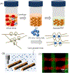
Schematic of the proposed patient-specific bioink. (a) Schematic of the extraction of PRP and its incorporation in alginate to form the bioink. (b) Schematic of the proposed bioprinting process. (c) A typical bioprinted scaffold from alginate-PRP containing fluorescent particles.
Figure 2.
Figure 2.
Physical characterizations of alginate/PRP hydrogels and assessment of protein release profile. (a) Compressive modulus of alginate (1% w/v) and alginate (1% w/v)/PRP (50U/mL) hydrogels. (b) Water uptake of Alginate and Alginate/PRP hydrogels in PBS-based solution after 24 hours. (c) Cumulative total protein release from alginate/PRP (50U/mL) hydrogel and PRP gel. (d) Cumulative VEGF release from alginate/PRP hydrogel (50U/mL). (**: P<0.01).
Figure 3.
Figure 3.
Effect of PRP-based bioink on the growth and function of MSCs. (a) Metabolic activity of MSC cells treated with alginate and alginate/PRP. (b, c) Scratch assay demonstrating the effect of PRP on migration and reorganization of MSCs in culture in comparison to the alginate as the negative control. (d) Schematic of migration assay using Boyden chamber and migration of MSC cells in the presence of chemo attractants released from alginate/PRP hydrogel. (*: P<0.05; **: P<0.01; ***: P<0.001)
Figure 4.
Figure 4.
(a) Metabolic activity of HUVECs treated with alginate and alginate/PRP over 3 days of culture in media without any growth factors; alginate is the negative control. (b) A typical tube formation assay for assessment of the function and morphology of HUVECs. HUVECs were cultured on collagen type I and treatment (alginate/PRP) was applied and the number of branch points and tube length were measured. (c, d) Summary of the total number of branch points measured in five fields under each condition. The micrographs confirmed a significant difference in cellular morphology. (*: P<0.05; **: P<0.01, ***: P<0.001)
Figure 5.
Figure 5.
3D bioprinting of the engineered bioink. (a-f) Images and micrographs of different constructs based on alginate and PRP. In the fabrication of these constructs a solution of 1% (w/v) alginate, 50 U/mL of PRP, and 0.04 % (w/v) CaCl2 was used. The digital files are also shown in (a,c,e). (g-i) 3D constructs could be detached from the substrate without losing their integrity and could be used as free standing constructs.

References

    1. Murphy SV, Atala A, Nature biotechnology 2014, 32, 773; - PubMed
    2. Mandrycky C, Wang Z, Kim K, Kim D-H, Biotechnology advances 2016, 34, 422. - PMC - PubMed
    1. Billiet T, Vandenhaute M, Schelfhout J, Van Vlierberghe S, Dubruel P, Biomaterials 2012, 33, 6020; - PubMed
    2. Liu W, Zhang YS, Heinrich MA, De Ferrari F, Jang HL, Bakht SM, Alvarez MM, Yang J, Li YC, Trujillo- de Santiago G, Advanced Materials 2017, 29. - PMC - PubMed
    1. Zhu K, Shin SR, van Kempen T, Li YC, Ponraj V, Nasajpour A, Mandla S, Hu N, Liu X, Leijten J, Advanced functional materials 2017, 27. - PMC - PubMed
    1. Ferris CJ, Gilmore KJ, Beirne S, McCallum D, Wallace GG, Biomaterials Science 2013, 1, 224. - PubMed
    1. Drury JL, Mooney DJ, Biomaterials 2003, 24, 4337; - PubMed
    2. Tibbitt MW, Anseth KS, Biotechnology and bioengineering 2009, 103, 655; - PMC - PubMed
    3. Annabi N, Tamayol A, Uquillas JA, Akbari M, Bertassoni LE, Cha C, Camci‐Unal G, Dokmeci MR, Peppas NA, Khademhosseini A, Advanced Materials 2014, 26, 85; - PMC - PubMed
    4. Fallahi A, Khademhosseini A, Tamayol A, Trends in biotechnology 2016, 34, 683. - PMC - PubMed

Publication types

MeSH terms