

Repair of a Large Ventral Hernia in a Rhesus Macaque (Macaca mulatta) by Using an Abdominal Component Separation Technique
- PMID: 29663944
- PMCID: PMC5897975
Repair of a Large Ventral Hernia in a Rhesus Macaque (Macaca mulatta) by Using an Abdominal Component Separation Technique
Abstract
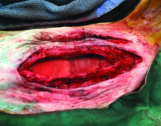
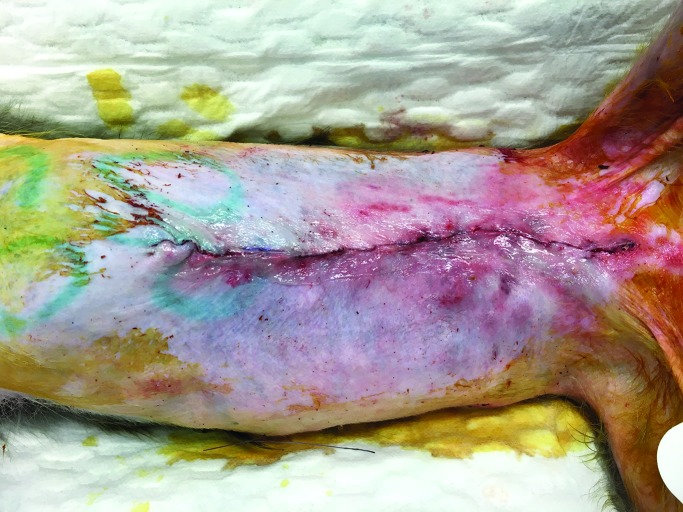
Here we present a 32-y-old rhesus macaque (Macaca mulatta) with a large recurrent ventral incisional hernia. The initial surgery included midline celiotomy for treatment of endometriosis, in which the animal developed a hernia that was repaired with interposition of mesh. Hernia recurrence at 1 y resulted in a defect measuring 7 × 13 cm, with loss of abdominal domain. Skin breakdown was noted with areas of exposed mesh through the skin with associated acute on chronic infection. Clinically, the animal was lethargic, not eating, and failing to thrive. The present surgical treatment included midline celiotomy, removal of mesh, and attempted primary fascial closure. Due to the large defect and high tension, the fascia could not be closed. To facilitate closure, abdominal component separation technique was used and consisted of skin and subcutaneous dissection, external oblique muscle release, and dissection between the external and internal oblique musculature. This technique allowed for primary fascial closure and resection of excess diseased skin. A piece of polypropylene mesh was placed in a sublay fashion to reinforce the primary fascial closure. The animal tolerated the procedure well and has demonstrated steady weight gain, with no recurrence at 12 mo. Large ventral abdominal hernia defects in after surgery or trauma in NHP can present reconstructive challenges to veterinary surgeons. Failure to achieve a dynamic, low-tension closure can result in hernia recurrence, necessitating additional operations. Abdominal component separation is not commonly used in veterinary surgery and may be a helpful tool in cases of difficult abdominal reconstructions.
Figures


References
-
- Afifi AM, Hartmann E, Talaat A, Alfotooh AA, Omar OS, Mareei S, Sanchez R, Kempton SJ. 2017. Quantitative assessment of tension reduction at the midline closure during abdominal component separation. J Am Coll Surg 224:954–961. - PubMed
-
- Albino FP, Patel KM, Nahabedian MY, Sosin M, Attinger CE, Bhanot P. 2013. Does mesh location matter in abdominal wall reconstruction? A systematic review of the literature and a summary of recommendations. Plast Reconstr Surg 132:1295–1304. - PubMed
-
- DiBello JN, Jr, Moore JH., Jr 1996. Sliding myofascial flap of the rectus abdominus muscles for the closure of recurrent ventral hernias. Plast Reconstr Surg 98:464–469. - PubMed
-
- Fry DE, Osler T. 1991. Abdominal wall considerations and complications in reoperative surgery. Surg Clin North Am 71:1–11. - PubMed
Publication types
MeSH terms
Grants and funding
LinkOut - more resources
Full Text Sources