

Predictors of vision impairment in Multiple Sclerosis
- PMID: 29664921
- PMCID: PMC5903642
- DOI: 10.1371/journal.pone.0195856
Predictors of vision impairment in Multiple Sclerosis
Abstract
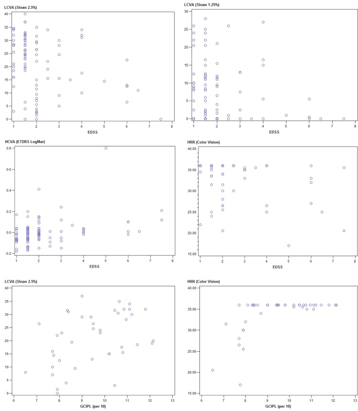
Visual impairment significantly alters the quality of life of people with Multiple Sclerosis (MS). The objective of this study was to identify predictors (independent variables) of visual outcomes, and to define their relationship with neurological disability and retinal atrophy when assessed by optical coherence tomography (OCT). We performed a cross-sectional analysis of 119 consecutive patients with MS, assessing vision using high contrast visual acuity (LogMar), 2.5% and 1.25% low contrast visual acuity (Sloan charts), and color vision (Hardy-Rand-Rittler plates). Quality of vision is a patient reported outcome based on an individual's unique perception of his or her vision and was assessed with the Visual Functioning Questionnaire-25 (VFQ-25) with the 10 neuro-ophthalmologic items. MS disability was assessed using the expanded disability status scale (EDSS), the MS functional composite (MSFC) and the brief repetitive battery-neuropsychology (BRB-N). Retinal atrophy was assessed using spectral domain OCT, measuring the thickness of the peripapillar retinal nerve fiber layer (pRNFL) and the volume of the ganglion cell plus inner plexiform layer (GCIPL). The vision of patients with MS was impaired, particularly in eyes with prior optic neuritis. Retinal atrophy (pRNFL and GCIPL) was closely associated with impaired low contrast vision and color vision, whereas the volume of the GCIPL showed a trend (p = 0.092) to be associated with quality of vision. Multiple regression analysis revealed that EDSS was an explanatory variable for high contrast vision after stepwise analysis, GCIPL volume for low contrast vision, and GCIPL volume and EDSS for color vision. The explanatory variables for quality of vision were high contrast vision and color vision. In summary, quality of vision in MS depends on the impairment of high contrast visual acuity and color vision due to the disease.
Conflict of interest statement
Figures

References
-
- Heesen C, Bohm J, Reich C, Kasper J, Goebel M, Gold SM. Patient perception of bodily functions in multiple sclerosis: gait and visual function are the most valuable. Mult Scler. 2008;14(7):988–91. doi: 10.1177/1352458508088916 . - DOI - PubMed
-
- Balcer LJ, Miller DH, Reingold SC, Cohen JA. Vision and vision-related outcome measures in multiple sclerosis. Brain. 2015;138(Pt 1):11–27. doi: 10.1093/brain/awu335 ; PubMed Central PMCID: PMCPMC4285195. - DOI - PMC - PubMed
-
- Rudick R, Antel J, Confavreux C, Cutter G, Ellison G, Fischer J, et al. Recommendations from the National Multiple Sclerosis Society Clinical Outcomes Assessment Task Force. Ann Neurol. 1997;42(3):379–82. doi: 10.1002/ana.410420318 - DOI - PubMed
-
- Balcer LJ, Baier ML, Pelak VS, Fox RJ, Shuwairi S, Galetta SL, et al. New low-contrast vision charts: reliability and test characteristics in patients with multiple sclerosis. Mult Scler. 2000;6(3):163–71. Epub 2000/06/29. doi: 10.1177/135245850000600305 . - DOI - PubMed
-
- Lampert EJ, Andorra M, Torres-Torres R, Ortiz-Perez S, Llufriu S, Sepulveda M, et al. Color vision impairment in multiple sclerosis points to retinal ganglion cell damage. J Neurol. 2015;262(11):2491–7. doi: 10.1007/s00415-015-7876-3 . - DOI - PubMed
Publication types
MeSH terms
LinkOut - more resources
Full Text Sources
Other Literature Sources
Medical
