. 2018 Jun 7;131(23):2581-2593.
doi: 10.1182/blood-2017-12-822619. Epub 2018 Apr 17.

Increased erythrophagocytosis induces ferroptosis in red pulp macrophages in a mouse model of transfusion

Affiliations

Increased erythrophagocytosis induces ferroptosis in red pulp macrophages in a mouse model of transfusion

Lyla A Youssef et al. Blood. .

Abstract

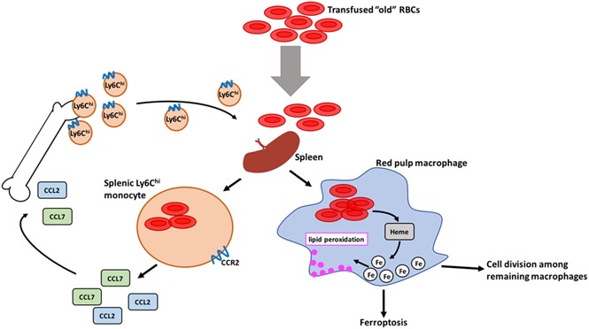
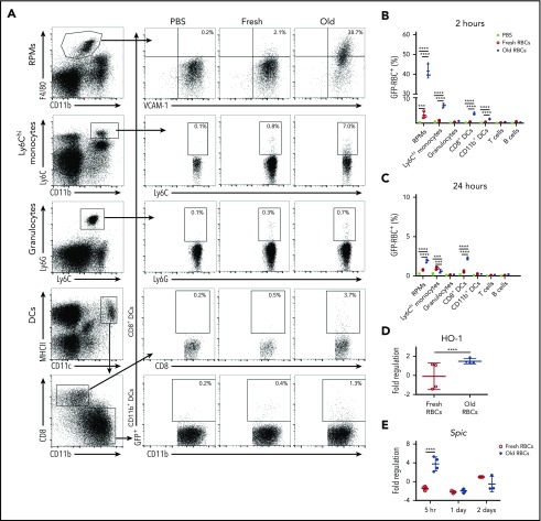
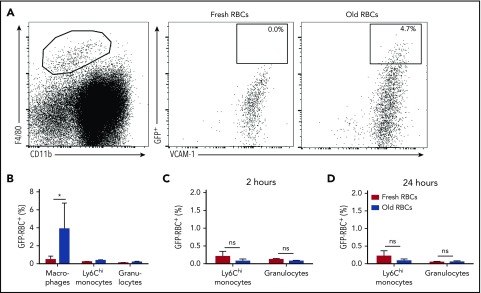
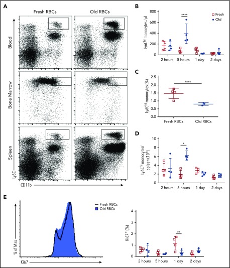
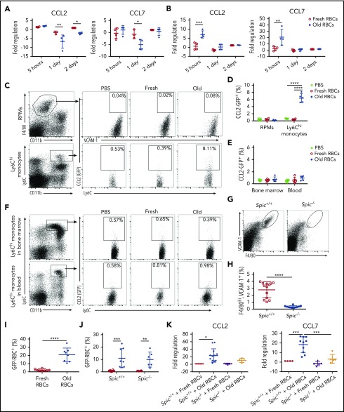
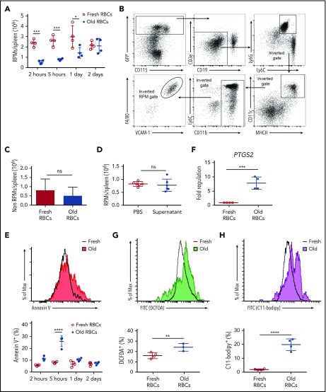
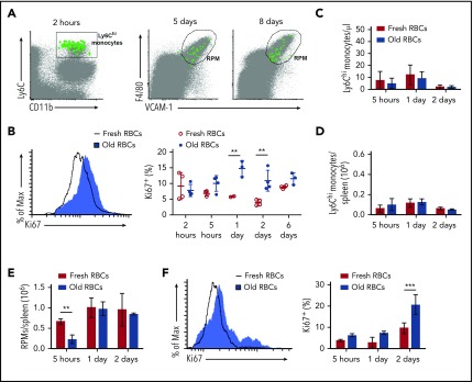
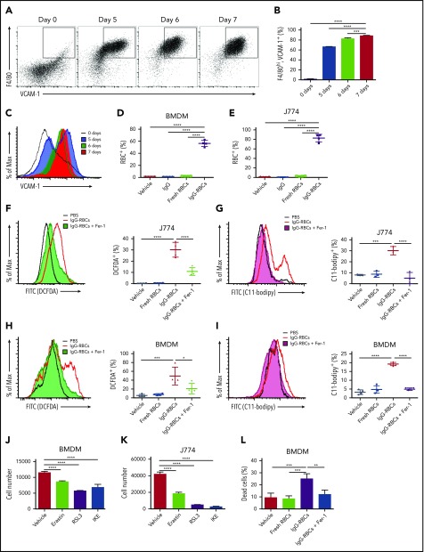
Macrophages play important roles in recycling iron derived from the clearance of red blood cells (RBCs). They are also a critically important component of host defense, protecting against invading pathogens. However, the effects on macrophage biology of acutely ingesting large numbers of RBCs are not completely understood. To investigate this issue, we used a mouse model of RBC transfusion and clearance, which mimics the clinical setting. In this model, transfusions of refrigerator storage-damaged (ie, "old") RBCs led to increased erythrophagocytosis by splenic red pulp macrophages (RPMs). This robust erythrophagocytosis induced ferroptosis, an iron-dependent form of cell death, in RPMs. This was accompanied by increases in reactive oxygen species and lipid peroxidation in vivo, which were reduced by treatment in vitro with ferrostatin-1, a ferroptosis inhibitor. Old RBC transfusions also induced RPM-dependent chemokine expression by splenic Ly6Chi monocytes, which signaled Ly6Chi monocyte migration from bone marrow to spleen, where these cells subsequently differentiated into RPMs. The combination of cell division among remaining splenic RPMs, along with the influx of bone marrow-derived Ly6Chi monocytes, suggests that, following RPM depletion induced by robust erythrophagocytosis, there is a coordinated effort to restore homeostasis of the RPM population by local self-maintenance and contributions from circulating monocytes. In conclusion, these findings may be clinically relevant to pathological conditions that can arise as a result of increased erythrophagocytosis, such as transfusion-related immunomodulation and impaired host immunity.


Conflict of interest statement

Conflict-of-interest disclosure: The authors declare no competing financial interests.

Figures

None
Graphical abstract
Figure 1.
Figure 1.
RPMs are the predominant splenic population responsible for phagocytosis of old RBCs, with Ly6Chimonocytes also contributing to RBC clearance. Wild-type C57BL/6 mice were transfused with 200 μL of PBS, fresh GFP+ RBCs, or old GFP+ RBCs. Mice were then sacrificed at 2 or 24 hours posttransfusion. (A) Representative dot plots show the gating strategy for RPMs (F4/80hi, VCAM-1+, CD11blo), Ly6Chi monocytes (Ly6Chi, CD11bhi), granulocytes (Ly6G+, Ly6C+, CD11b+), CD8+ DCs (CD8+, CD11b, MHCIIhi, CD11chi), and CD11b+ DCs (CD8, CD11b+, MHCIIhi, CD11chi) at 2 hours posttransfusion. These cells were obtained from the spleen at 2 hours posttransfusion. Percentages of GFP+ cells are depicted; these represent nucleated cells that have phagocytosed GFP-expressing fresh or old transfused RBCs. (B) Frequency of GFP+ RBC phagocytosis at 2 and (C) 24 hours posttransfusion; by 24 hours posttransfusion, the levels of phagocytosis were dramatically reduced compared with the 2-hour time point and the previously ingested GFP+ RBCs were degraded. (D) Hmox-1 (ie, HO-1) expression by RPMs 5 hours posttransfusion. (E) Spic expression by Ly6Chi monocytes 5 hours posttransfusion. Data representative of 4 experiments with at least 3 mice per group; *P < .05; ***P < .001; ****P < .0001; ANOVA with Tukey or Sidak multiple comparison test.
Figure 2.
Figure 2.
VCAM-1+, F4/80hibone marrow macrophages ingest transfused old RBCs. (A) Gating strategy for identifying bone marrow macrophages (VCAM-1+, F4/80hi, CD11blo) that phagocytose transfused RBCs (GFP+). (B) Frequency of phagocytosis of old RBCs at 2 hours posttransfusion by bone marrow macrophages, Ly6Chi monocytes and granulocytes; although macrophages ingest some transfused RBCs, no such phagocytosis is seen with Ly6Chi monocytes or granulocytes. (C-D) Frequency of ingested GFP+ RBCs at 2 and 24 hours posttransfusion in circulating Ly6Chi monocytes and granulocytes. Data representative of 3 experiments with at least 3 mice per group. *P < .05; ANOVA with Tukey or Sidak multiple comparison test. ns, not significant.
Figure 3.
Figure 3.
Ly6Chimonocytes emigrate from the bone marrow and home to the spleen after transfusion of old RBCs. Wild-type C57BL/6 mice were transfused with 350 μL of fresh or old RBCs and sacrificed at defined time points. (A) Flow plots depict increases in Ly6Chi monocytes in the blood and spleen and a decrease in the bone marrow at 5 hours posttransfusion. (B) Quantification of circulating Ly6Chi monocytes at defined time points demonstrating increased levels at 5 hours posttransfusion. (C) Quantification of bone marrow Ly6Chi monocytes demonstrating decreased levels at 5 hours posttransfusion. (D) Splenic Ly6Chi monocyte levels increase substantially at 5 hours posttransfusion. (E) Histogram of Ki67 staining of splenic Ly6Chi monocytes at 5 hours posttransfusion. Data representative of 3 experiments with at least 3 mice per group. *P < .05; **P < .01; ****P < .0001; ANOVA followed by Tukey posttest.
Figure 4.
Figure 4.
CCL2 and CCL7 mRNA expression by splenic Ly6Chimonocytes following enhanced erythrophagocytosis requires the presence of RPMs. At defined time points after transfusing wild-type recipients with 350 μL of fresh or old RBCs, splenocytes were isolated by FACS, and CCL2 and CCL7 mRNA expression was measured by qPCR in (A) RPMs and (B) Ly6Chi monocytes; these data show that, after old RBC transfusions, expression of these chemokines did not change, or even decreased, in RPMs, whereas they both increased substantially in Ly6Chi monocytes at 5 hours posttransfusion. CCL2-GFP reporter mice were transfused with 200 μL of PBS, fresh GFP+ RBCs, or old GFP+ RBCs, and then sacrificed at 2 hours posttransfusion. (C) Gating strategy for splenic RPMs and Ly6Chi monocytes along with representative dot plots showing induced CCL2 promoter–dependent GFP expression; the percentages of cells expressing GFP are provided. (D) Frequency of GFP expression, demonstrating upregulation by Ly6Chi monocytes, but not by RPMs. (E) Frequency of CCL2 expression by RPMs and Ly6Chi monocytes showing no statistically significant changes posttransfusion. (F) Gating strategy for Ly6Chi monocytes in the blood and bone marrow, along with representative dot plots showing CCL2 promoter–dependent GFP expression; the percentages of cells expressing GFP are provided. To determine the dependence of Ly6Chi monocyte CCL2 and CCL7 mRNA expression on the presence of RPMs, Spic−/− mice were used for the experiments in panels G-K. (G-H) RPMs (VCAM-1hi, F4/80hi) are absent in Spic−/− mice, as compared with Spic+/+ controls. (I) The proportion of RPMs in Spic+/+ recipient mice that ingested old GFP+ RBCs. (J) The proportions of Ly6Chi monocytes in Spic+/+ and Spic−/− mice that ingested old GFP+ RBCs. (K) Fold regulation of CCL2 and CCL7 mRNA expression, as measured by qPCR, in splenic Ly6Chi monocytes from Spic+/+ and Spic−/− mice. *P < .05; **P < .01; ***P < .001; ****P < .0001; ANOVA with Sidak or Tukey multiple comparisons test or unpaired t test. Data are representative of 3 independent experiments, except for Spic+/+ and Spic−/− results, which were pooled for all experiments.
Figure 5.
Figure 5.
Transfusions of old RBCs induce oxidative stress, PTGS2 expression, and cell death in RPMs. (A) Wild-type C57BL/6 mice were transfused with 350 μL of fresh or old RBCs and sacrificed at defined time points. Splenic RPM cell numbers were quantified posttransfusion. Old RBC transfusions induced a substantial decrease in RPMs by 2 hours posttransfusion, the levels of which returned to baseline 2 days later. (B) The gating strategy first selects for splenic nucleated non-RPM cells, which thereby selects for GFP+ (Csfr1/CD115+) cells; this is followed by gating out B cells, granulocytes, DCs, monocytes, and RPMs. The remaining cells were quantified to determine changes following transfusions of old, as compared with fresh, RBCs. (C) MaFIA mice were transfused with 350 μL of fresh or old RBCs and sacrificed 5 hours posttransfusion. Total non-RPM–nucleated cell numbers in the spleen were quantified, showing no differences. (D) Wild-type C57BL/6 mice were infused with PBS or supernatant from 350 μL of an old RBC blood bank and RPM cell number was quantified 5 hours posttransfusion. (E) Annexin V staining of RPMs following transfusion of old and fresh RBCs. (F) Upregulation of PTGS2 mRNA expression in RPMs after old RBC transfusions. Increases in ROS production (G) and lipid peroxidation (H) in RPMs following transfusions of old RBCs. *P < .05; **P < .01; ***P < .001; ****P < .0001; ANOVA with the Sidak multiple comparisons test. PTGS2 gene expression analysis was analyzed for significance using the unpaired Student t test. Data are representative of 3 independent experiments. FITC, fluorescein isothiocyanate.
Figure 6.
Figure 6.
Splenic RPMs exhibit local self-maintenance following old RBC transfusion-induced cell death. (A) Bone marrow from UBC-GFP mice was harvested and enriched for Ly6Chi monocytes by negative selection. GFP+, Ly6Chi, CD115+, CD11b+ monocytes were then adoptively transferred to wild-type C57BL/6 recipients prior to transfusion of the latter with 350 μL of old RBCs. At 2 hours posttransfusion, GFP+ Ly6Chi monocytes were observed in the spleen. By 5 and 8 days posttransfusion, GFP+ RPMs (F4/80hi, VCAM-1hi) were observed in the spleen. (B) Wild-type C57BL/6 mice were transfused with 350 μL of fresh or old RBCs and sacrificed at defined time points. Splenic RPMs were stained with Ki67, demonstrating increased proliferation at 1 to 2 days after old RBC transfusions, as compared with RPMs from mice that received fresh RBCs. Histogram (left) of Ki67 staining at 1 day posttransfusion. (C) Ly6Chi monocyte cell numbers were quantified in the blood of CCR2−/− mice, demonstrating their absence. (D) Ly6Chi monocyte cell numbers were quantified in the spleens of CCR2−/− mice, demonstrating their absence. (E) The splenic RPM population in CCR2−/− mice is reduced by 5 hours after transfusions of old RBCs, but restored by 2 days posttransfusion. (F) By Ki67 staining, RPMs in CCR2−/− mice proliferate following transfusion of old RBCs, relative to RPMs in mice that received fresh RBCs. Histogram (left) of Ki67 at 2 days posttransfusion. **P < .01; ***P < .001; ANOVA with the Sidak posttest or unpaired Student t test. Data are representative of 3 independent experiments.
Figure 7.
Figure 7.
Enhanced erythrophagocytosis induces ferroptosis in macrophages in vitro. Bone marrow was harvested from wild-type C57BL/6 mice and cultured in the presence of macrophage colony-stimulating factor (20 ng/mL) for defined times to determine when the cell population uniformly expressed VCAM-1 and F4/80. (A) Representative dot plots (the percentages of double-positive cells are indicated), (B) frequency of VCAM-1+, F4/80hi macrophages, and (C) overall VCAM-1 expression are shown. BMDMs and J774 cells were plated 24 hours prior to the experiments and then incubated with PBS, fresh RBCs, or IgG-opsonized RBCs, which were labeled with CellTrace Far Red. The amount of erythrophagocytosis in (D) BMDMs and (E) J774 cells was quantified by flow cytometry. J774 cells (F-G) and BMDMs (H-I) were treated with vehicle control or 100 μM Fer-1 and exposed to IgG-opsonized RBCs; they were then analyzed for ROS (F,H) and lipid peroxidation (G,I). Twenty-four-hour exposure to erastin (20 μM), RSL3 (1 μM), and IKE (20 μM), which are potent ferroptosis inducers, induced BMDM (J) and J774 cell loss (K). BMDM cell viability was quantified following incubation with IgG-opsonized RBCs or PBS, and with vehicle control or 100 μM Fer-1 for 6 hours; enhanced erythrophagocytosis induced substantial cell death, which was ameliorated by Fer-1 (L). Data are representative of 3 independent experiments. *P < .05; ***P < .001; ****P < .0001; ANOVA with the Dunnett multiple comparisons test.

References

    1. Smith LP, Hunter KW, Oldfield EC, Strickland GT. Murine malaria: blood clearance and organ sequestration of Plasmodium yoelii-infected erythrocytes. Infect Immun. 1982;38(1):162-167. - PMC - PubMed
    1. Strobel E. Hemolytic transfusion reactions. Transfus Med Hemother. 2008;35(5):346-353. - PMC - PubMed
    1. Chaudhary RK, Das SS. Autoimmune hemolytic anemia: from lab to bedside. Asian J Transfus Sci. 2014;8(1):5-12. - PMC - PubMed
    1. Luzzatto L, Nannelli C, Notaro R. Glucose-6-phosphate dehydrogenase deficiency. Hematol Oncol Clin North Am. 2016;30(2):373-393. - PubMed
    1. Hod EA, Zhang N, Sokol SA, et al. Transfusion of red blood cells after prolonged storage produces harmful effects that are mediated by iron and inflammation. Blood. 2010;115(21):4284-4292. - PMC - PubMed

Publication types