Review
. 2018 May 7;150(5):671-682.
doi: 10.1085/jgp.201711980. Epub 2018 Apr 17.

The ins and outs of vesicular monoamine transporters

Affiliations
Review

The ins and outs of vesicular monoamine transporters

Dana Yaffe et al. J Gen Physiol. .

Abstract

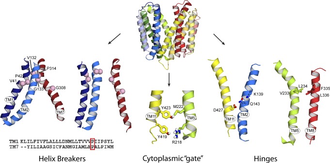
The H+-coupled vesicular monoamine transporter (VMAT) is a transporter essential for life. VMAT mediates packaging of the monoamines serotonin, dopamine, norepinephrine, and histamine from the neuronal cytoplasm into presynaptic vesicles, which is a key step in the regulated release of neurotransmitters. However, a detailed understanding of the mechanism of VMAT function has been limited by the lack of availability of high-resolution structural data. In recent years, a series of studies guided by homology models has revealed significant insights into VMAT function, identifying residues that contribute to the binding site and to specific steps in the transport cycle. Moreover, to characterize the conformational transitions that occur upon binding of the substrate and coupling ion, we have taken advantage of the unique and powerful pharmacology of VMAT as well as of mutants that affect the conformational equilibrium of the protein and shift it toward defined conformations. This has allowed us to identify an important role for the proton gradient in driving a shift from lumen-facing to cytoplasm-facing conformations.


Figures

Figure 1.
Figure 1.
Membrane-embedded charged residues in VMAT2. Top: Structural model of rVMAT2 in the lumen-facing conformation (Clum’), indicating the position of membrane-embedded charged residues in rVMAT2 (spheres) and colored according to TM helix, with the N-terminal half in shades of blue and green, and the C-terminal half in shades of red and yellow. Bottom: TM topology of antiporters from the DHA-1 family, colored according to the structure figure above. The structure of YajR (Protein Data Bank accession no. 3WDO) and the homology model of BbMAT were aligned to identify the conserved membrane-embedded charged residues, which are indicated as ellipses, based on the color coding indicated in the legend. Transparent triangles indicate the location of three-helix structural repeats in the N-terminal (blue and green) and C-terminal halves (pink and yellow) of the MFS fold.
Figure 2.
Figure 2.
Structural elements involved in conformational changes during the rVMAT2 transport cycle. Top: Model of rVMAT2 in a lumen-facing state (Clum’) viewed along the plane of the membrane with the lumen at the top. Bottom left: Helix breakers. TM helices 1, 2, and 7 in the lumen-facing (left) and cytoplasm-facing (right) conformations are shown as cartoon helices, and relevant glycine and proline residues are indicated using pink spheres. Sequence alignment of helices 1 and 7. Helices were defined using the Clum’ model and aligned using ClustalW. Bottom middle: Gating residues. Magnification of the cytoplasmic domain of TM5 and TM11. Residues contributing to the cytoplasmic gate are shown as sticks. Bottom right: Hinges. Residues predicted to form interactions between TM2 and TM11 and between TM5 and TM8 in the vesicle lumen-facing conformation of rVMAT2 (sticks). These residues were predicted to form hinge points for the conformational change because the relative position of these residues is essentially unchanged between the cytoplasm and vesicle lumen-facing models. Adapted from Ugolev et al. (2013) and Yaffe et al.2013, 2016).
Figure 3.
Figure 3.
Schematic of the proposed transport cycle. For simplicity, only six TMs are shown. The cycle is assumed to involve eight steps (numbered). In the absence of a proton gradient, the dominant population is of the lumen-facing conformation, as indicated by the transporter’s ability to bind tetrabenazine but not reserpine. Binding of protons enables the conformational switch to the cytoplasm-facing conformation (step 1), whereas binding of substrate enables the change to the lumen-facing conformation (step 5). Binding of tetrabenazine locks the transporter in a conformation that appears incompatible with substrate binding and is therefore presumably not cytoplasm facing (shown as an off-cycle state connected to step 8). Binding of reserpine also locks the transporter in a dead-end conformation, but reserpine binding competes with substrate binding, and therefore the reserpine-bound conformation is presumably cytoplasm facing (shown as an off-cycle state connected to step 4). The dashed arrow for reserpine indicates competition with substrate. Adapted from Yaffe et al. (2016).

References

    1. Abramson J., Smirnova I., Kasho V., Verner G., Kaback H.R., and Iwata S.. 2003. Structure and mechanism of the lactose permease of Escherichia coli. Science. 301:610–615. 10.1126/science.1088196 - DOI - PubMed
    1. Adler J., and Bibi E.. 2004. Determinants of substrate recognition by the Escherichia coli multidrug transporter MdfA identified on both sides of the membrane. J. Biol. Chem. 279:8957–8965. 10.1074/jbc.M313422200 - DOI - PubMed
    1. Adler J., Lewinson O., and Bibi E.. 2004. Role of a conserved membrane-embedded acidic residue in the multidrug transporter MdfA. Biochemistry. 43:518–525. 10.1021/bi035485t - DOI - PubMed
    1. Anne C., and Gasnier B.. 2014. Vesicular neurotransmitter transporters: mechanistic aspects. Curr. Top. Membr. 73:149–174. 10.1016/B978-0-12-800223-0.00003-7 - DOI - PubMed
    1. Buchanan B.B., Eiermann W., Riccio P., Aquila H., and Klingenberg M.. 1976. Antibody evidence for different conformational states of ADP, ATP translocator protein isolated from mitochondria. Proc. Natl. Acad. Sci. USA. 73:2280–2284. 10.1073/pnas.73.7.2280 - DOI - PMC - PubMed

Publication types

MeSH terms

Substances

LinkOut - more resources