Review
. 2018 May 7;215(5):1273-1285.
doi: 10.1084/jem.20180325. Epub 2018 Apr 17.

Type I interferons in tuberculosis: Foe and occasionally friend

Affiliations
Review

Type I interferons in tuberculosis: Foe and occasionally friend

Lúcia Moreira-Teixeira et al. J Exp Med. .

Abstract

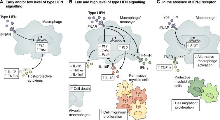
Tuberculosis remains one of the leading causes of mortality worldwide, and, despite its clinical significance, there are still significant gaps in our understanding of pathogenic and protective mechanisms triggered by Mycobacterium tuberculosis infection. Type I interferons (IFN) regulate a broad family of genes that either stimulate or inhibit immune function, having both host-protective and detrimental effects, and exhibit well-characterized antiviral activity. Transcriptional studies have uncovered a potential deleterious role for type I IFN in active tuberculosis. Since then, additional studies in human tuberculosis and experimental mouse models of M. tuberculosis infection support the concept that type I IFN promotes both bacterial expansion and disease pathogenesis. More recently, studies in a different setting have suggested a putative protective role for type I IFN. In this study, we discuss the mechanistic and contextual factors that determine the detrimental versus beneficial outcomes of type I IFN induction during M. tuberculosis infection, from human disease to experimental mouse models of tuberculosis.


Figures

Figure 1.
Figure 1.
Alternative pathways of type I IFN induction during M. tuberculosis infection. Recognition of mycobacterial products by a range of cell surface and cytosolic PRR, including TLR4, NOD2, and STING, activates the kinase TBK1 leading to phosphorylation (P) and dimerization of IRF3 or IRF5, which translocates into the nucleus and promotes transcription of type I IFN genes. Release of mycobacterial or mitochondrial DNA in the cytosol activates cGAS, which synthesizes cGAMP. Host-derived cGAMP and/or mycobacterial-derived c-di-AMP activates the STING pathway and the downstream TBK1–IRF3 signaling axis. Peptidoglycan fragments can be sensed by NOD2 in the cytosol, activating the TBK1–IRF5 signaling pathway. Detection of extracellular M. tuberculosis and/or its products by TLR4 triggers TRIF-TBK1-IRF3–dependent induction of type I IFN by certain strains. Mtb, M. tuberculosis; mtDNA, mitochondrial DNA; PGN, peptidoglycan.
Figure 2.
Figure 2.
Foe- and friendly-like effects of type I IFN during M. tuberculosis infection. Type I IFN has been reported to play both negative (red arrows) and positive (green arrows) functions during M. tuberculosis infection. (A) Tonic levels of autocrine type I IFN signaling prime the production of protective cytokines IL-12 and TNF-α. (B) However, high and sustained levels of type I IFN promote the production of IL-10 and inhibit the production of protective cytokines IL-12, TNF-α, IL-1α, and IL-1β. IL-10 mediates a suppressive feedback loop, contributing to the decreased production of IL-12 and TNF-α. Type I IFN also inhibits myeloid cell responsiveness to IFN-γ by both IL-10–dependent and independent mechanisms, suppressing IFN-γ–dependent host-protective immune responses. In addition, type I IFN can promote cell death in alveolar macrophages and accumulation of permissive myeloid cells at the site of infection. (C) In the absence of the IFN-γ receptor, type I IFN inhibits Arg1 expression directly or indirectly by increasing TNF-α levels, thus regulating macrophage activation toward a more protective phenotype. Type I IFN signaling can also promote the recruitment, differentiation, and/or survival of protective myeloid cells that control pathology at the site of infection. Arg1, arginase 1; IFNγR, IFN-γ receptor; IL-10R, IL-10 receptor; TNFR, TNF-α receptor.

References

    1. Abutidze A., Bolokadze N., Chkhartishvili N., Sharvadze L., and Tsertsvadze T.. 2016. Incidence of Tuberculosis among Hiv/Hcv Co-Infected Patients Receiving Hepatitis C Treatment with Pegylated Interferon and Ribavirin in Georgia. Georgian Med. News. 252:10–15. - PMC - PubMed
    1. Altare F., Durandy A., Lammas D., Emile J.F., Lamhamedi S., Le Deist F., Drysdale P., Jouanguy E., Döffinger R., Bernaudin F., et al. . 1998. Impairment of mycobacterial immunity in human interleukin-12 receptor deficiency. Science. 280:1432–1435. 10.1126/science.280.5368.1432 - DOI - PubMed
    1. Anderson S.T., Kaforou M., Brent A.J., Wright V.J., Banwell C.M., Chagaluka G., Crampin A.C., Dockrell H.M., French N., Hamilton M.S., et al. . 2014. Diagnosis of childhood tuberculosis and host RNA expression in Africa. N. Engl. J. Med. 370:1712–1723. 10.1056/NEJMoa1303657 - DOI - PMC - PubMed
    1. Antonelli L.R., Gigliotti Rothfuchs A., Gonçalves R., Roffê E., Cheever A.W., Bafica A., Salazar A.M., Feng C.G., and Sher A.. 2010. Intranasal Poly-IC treatment exacerbates tuberculosis in mice through the pulmonary recruitment of a pathogen-permissive monocyte/macrophage population. J. Clin. Invest. 120:1674–1682. 10.1172/JCI40817 - DOI - PMC - PubMed
    1. Banchereau J., and Pascual V.. 2006. Type I interferon in systemic lupus erythematosus and other autoimmune diseases. Immunity. 25:383–392. 10.1016/j.immuni.2006.08.010 - DOI - PubMed

Publication types

Substances