. 2018 Jun 15;293(24):9265-9276.
doi: 10.1074/jbc.RA117.000365. Epub 2018 Apr 17.

Lmo1656 is a secreted virulence factor of Listeria monocytogenes that interacts with the sorting nexin 6-BAR complex

Affiliations

Lmo1656 is a secreted virulence factor of Listeria monocytogenes that interacts with the sorting nexin 6-BAR complex

Daryl Jason David et al. J Biol Chem. .

Abstract

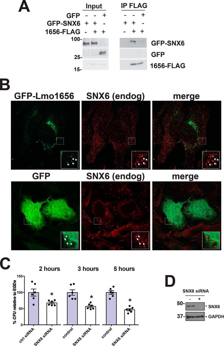
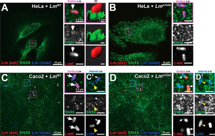
Listeria monocytogenes (Lm) is a facultative intracellular bacterial pathogen and the causative agent of listeriosis, a rare but fatal disease. During infection, Lm can traverse several physiological barriers; it can cross the intestine and placenta barrier and, in immunocompromised individuals, the blood-brain barrier. With the recent plethora of sequenced genomes available for Lm, it is clear that the complete repertoire of genes used by Lm to interact with its host remains to be fully explored. Recently, we focused on secreted Lm proteins because they are likely to interact with host cell components. Here, we investigated a putatively secreted protein of Lm, Lmo1656, that is present in most sequenced strains of Lm but absent in the nonpathogenic species Listeria innocua. lmo1656 gene is predicted to encode a small, positively charged protein. We show that Lmo1656 is secreted by Lm Furthermore, deletion of the lmo1656 gene (Δlmo1656) attenuates virulence in mice infected orally but not intravenously, suggesting that Lmo1656 plays a role during oral listeriosis. We identified sorting nexin 6 (SNX6), an endosomal sorting component and BAR domain-containing protein, as a host cell interactor of Lmol656. SNX6 colocalizes with WT Lm during the early steps of infection. This colocalization depends on Lmo1656, and RNAi of SNX6 impairs infection in infected tissue culture cells, suggesting that SNX6 is utilized by Lm during infection. Our results reveal that Lmo1656 is a novel secreted virulence factor of Lm that facilitates recruitment of a specific member of the sorting nexin family in the mammalian host.

Keywords: Listeria monocytogenes; Lmo1656; bacterial pathogenesis; host-pathogen interaction; infection; sorting nexin (SNX); sorting nexin 6; virulence factor.


Conflict of interest statement

The authors declare that they have no conflicts of interest with the contents of this article

Figures

Figure 1.
Figure 1.
Lmo1656 is a predicted secreted protein of L. monocytogenes. A, synteny of the lmo1656 locus. lmo1656 is conserved in most sequenced strains of L. monocytogenes but absent in the closely related but nonpathogenic L. innocua. Epidemic Lm strain F2365 is shown as an example of a clinical isolate. B, homologs of lmo1656 are predicted in other bacterial species, most of which are Gram-positive. Multiple sequence alignment (ClustalX2) of the predicted proteins, excluding the putative Sec-dependent signal peptide. The mature form of Lmo1656 is predicted to have a molecular mass of 12.49 kDa and a pI of 10.61.
Figure 2.
Figure 2.
Lmo1656 is a secreted protein. A, overexpressed Lmo1656-FLAG is secreted into the growth medium. Either WT (LmWT) or Lmo1656-FLAG-overexpressing (Lm1656-FL+) bacteria were grown to exponential phase in broth medium. The sterile filtered supernatant was immunoprecipitated against FLAG. Lmo1656-FLAG is immunoprecipitated in the sterile filtered supernatant of the growth medium from Lm1656-FL+ but not LmWT bacteria. Data are representative of three independent experiments (Pt, bacterial pellet; In, input fraction; IP, immunoprecipitated fraction). B, secreted Lmo1656-FLAG can be immunoprecipitated by antibodies raised against Lmo1656. Either WT (LmWT), lmo1656 deletion mutant (LmΔ1656), or Lmo1656-FLAG–overexpressing (Lm1656-FL+) bacteria were grown to exponential phase in broth medium. The sterile filtered supernatants were immunoprecipitated with a pooled mixture of affinity-purified anti-Lmo1656 polyclonal rabbit antibodies (1 μg of antibody/50 μg of protein). Samples were subjected to Western blotting with the same anti-Lmo1656 antibody pool (1:500 in TBS, Tween 20, and 5% milk) (Pt, bacterial pellet; In, input fraction; Un, unbound fraction postimmunoprecipitation; IP, immunoprecipitated fraction). C, overexpressed Lmo1656-FLAG is secreted into infected cells. JEG3 cells were infected (m.o.i., 20) with either WT (LmWT) or Lmo1656-FLAG–overexpressing (Lm1656-FL+) bacteria. The cells were lysed 4 hpi and subjected to immunoprecipitation against FLAG. Lmo1656-FLAG is immunoprecipitated in the soluble lysate from cells infected with Lm1656-FL+ but not LmWT bacteria. Data are representative of three independent experiments (Pt, insoluble pellet postlysis; In, input fraction; IP, immunoprecipitated fraction). D, overexpressed Lmo1656-GFP localizes to endomembranes. HeLa cells were transiently transfected with Lmo1656-GFP for 24 h. Actin and nuclei were stained with phalloidin and DAPI, respectively. Images were acquired with a spinning disk confocal microscope. Data are representative of at least three independent experiments.
Figure 3.
Figure 3.
Lmo1656 is a bona fide virulence factor of L. monocytogenes. A and B, Lmo1656 contributes to early infection in certain cell types. LmΔlmo1656 have decreased bacteria at early time points (A; t = 2 hpi) but not later (B; t = 4 or 24 hpi) of infection in HeLa but not Caco2 or mouse bone marrow–derived macrophages (BMDM). Results are normalized to the mean cfu for WT per replicate (n = 3 wells per replicate over at least two independent replicates; *, p = 0.0339, analysis of variance). C and D, Lmo1656 contributes to oral infection in vivo. Knockin mice expressing a “humanized” E-CadE16P were infected with LmWT, LmΔlmo1656, or the complemented LmΔlmo1656+C via oral gavage. Mouse livers have decreased bacterial burden both 48 (C) and 72 hpi (D) when infected by LmΔlmo1656, whereas spleens have decreased bacterial burden 72 hpi (D) (n = 7–8 mice per Lm genotype; *, p = 0.0261 Mann–Whitney U test; data are means ± S.D. of at least three independent experiments).
Figure 4.
Figure 4.
SNX6 and Lmo1656 interact biochemically and genetically. A, GFP-SNX6 and Lmo1656-FLAG biochemically interact. HeLa cells were transiently transfected with GFP-SNX6 or GFP and either empty vector (−) or Lmo1656-FLAG (+). Anti-FLAG immunoprecipitation (IP) was performed on clarified lysate 48 h post-transfection. GFP-SNX6 can be coimmunoprecipitated with Lmo1656-FLAG from cells transfected with Lmo1656-FLAG but not with empty vector. Results are representative of two independent experiments. B, Lmo1656-GFP colocalizes with SNX6. HeLa cells were transiently transfected with Lmo1656-GFP (upper panel) or GFP (lower panel) and stained for endogenous (endog) SNX6. Inset, magnification of colocalizing Lmo1656-GFP and SNX6. C, SNX6 contributes to Lm infection. HeLa cells were transiently transfected with either nontargeting siRNA pool (control (ctrl)) or an siRNA pool targeting SNX6. 72 h post-transfection, cells were infected with Lm EGDe-PrfA* and lysed 2, 3, and 5 hpi. (*, p < 0.05; data are means ± S.D. of at least three independent experiments). Results are in triplicate from two independent experiments. D, knockdown of SNX6 in HeLa cells. HeLa cells were either treated with scrambled siRNA (−) or siRNA against SNX6 (+). After 72h, protein levels were analyzed by Western blotting with the indicated antibodies.
Figure 5.
Figure 5.
SNX6 colocalizes with Lm entry sites in an Lmo1656-dependent manner. A and B, endogenous SNX6 is recruited to internalizing Lm in an Lmo1656-dependent manner in HeLa cells. HeLa cells infected with either LmWT (A) or LmΔ1656 (B) (m.o.i., 40; 2 hpi) and stained for external (ext) and internalized bacteria. A, right, 3D surface reconstruction of endogenous SNX6 recruited to LmWT entry sites (Imaris). C and D, endogenous SNX6 is recruited to internalizing Lm in an Lmo1656-independent manner in Caco2 cells. Caco2 cells were infected with either LmWT (C) or LmΔ1656 (D) (m.o.i., 10; 2 hpi) and stained for external bacteria. Internalized (int) LmWT (C′) or LmΔ1656 (D′) Lm colocalize with SNX6. Magnification of bacteria (purple squares) is shown in adjacent images. Data are representative of at least three independent experiments; controls (LmWT or LmΔ1656) for each cell type were imaged with spinning disk confocal microscope during the same session using identical settings and, where necessary, identical adjustments for brightness and contrast. (A and B versus C and D).
Figure 6.
Figure 6.
Certain GFP-SNX–BAR proteins are recruited to Lm entry sites in an Lmo1656-dependent manner. A–C, Lm entry sites do not recruit a subset of GFP-SNX proteins. HeLa cells were transiently transfected with GFP-SNX constructs and stained for external (ex) Lm (m.o.i., 20; 2 hpi). A, GFP-SNX1; B, GFP-SNX2; C, GFP-SNX3. D and E, Lm entry sites recruit some GFP-SNX–BAR family proteins. HeLa cells were transiently transfected with GFP-SNX–BAR constructs and stained for external (ext) LmWT. D and D′, GFP-SNX5 with LmWT or LmΔ1656, respectively; E and E′, GFP-SNX6 with LmWT or LmΔ1656, respectively. Magnification of bacteria (purple squares) is shown in adjacent images. Data are representative of at least three independent experiments; controls (LmWT or LmΔ1656) were imaged with a spinning disk confocal microscope during the same session using identical settings and, where necessary, identical adjustments for brightness and contrast. tot, total.

References

    1. Ribet, D., and Cossart, P. (2015) How bacterial pathogens colonize their hosts and invade deeper tissues. Microbes Infect. 17, 173–183 10.1016/j.micinf.2015.01.004 - DOI - PubMed
    1. Pizarro-Cerdá, J., Kühbacher, A., and Cossart, P. (2012) Entry of Listeria monocytogenes in mammalian epithelial cells: an updated view. Cold Spring Harb. Perspect. Med. 2, a010009 10.1101/cshperspect.a010009 - DOI - PMC - PubMed
    1. Radoshevich, L., and Dussurget, O. (2016) Cytosolic innate immune sensing and signaling upon infection. Front. Microbiol. 7, 313 10.3389/fmicb.2016.00313 - DOI - PMC - PubMed
    1. de Souza Santos, M., and Orth, K. (2015) Subversion of the cytoskeleton by intracellular bacteria: lessons from Listeria, Salmonella and Vibrio. Cell. Microbiol. 17, 164–173 10.1111/cmi.12399 - DOI - PMC - PubMed
    1. Welch, M. D., and Way, M. (2013) Arp2/3-mediated actin-based motility: a tail of pathogen abuse. Cell Host Microbe 14, 242–255 10.1016/j.chom.2013.08.011 - DOI - PMC - PubMed

Publication types