. 2018 May 8;115(19):E4350-E4357.
doi: 10.1073/pnas.1718804115. Epub 2018 Apr 17.

Characterization and engineering of a plastic-degrading aromatic polyesterase

Affiliations

Characterization and engineering of a plastic-degrading aromatic polyesterase

Harry P Austin et al. Proc Natl Acad Sci U S A. .

Abstract

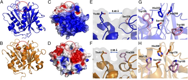
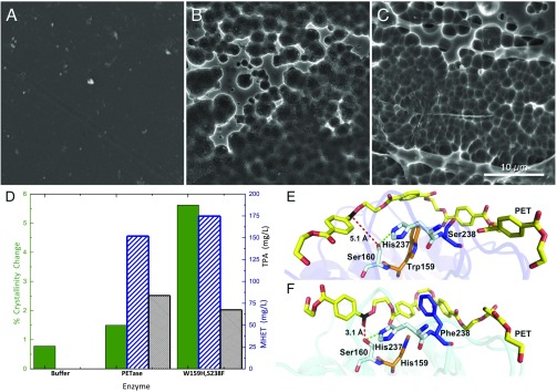
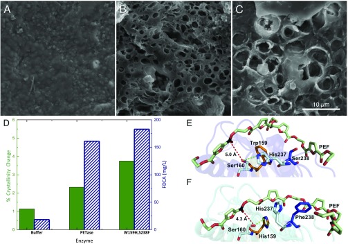
Poly(ethylene terephthalate) (PET) is one of the most abundantly produced synthetic polymers and is accumulating in the environment at a staggering rate as discarded packaging and textiles. The properties that make PET so useful also endow it with an alarming resistance to biodegradation, likely lasting centuries in the environment. Our collective reliance on PET and other plastics means that this buildup will continue unless solutions are found. Recently, a newly discovered bacterium, Ideonella sakaiensis 201-F6, was shown to exhibit the rare ability to grow on PET as a major carbon and energy source. Central to its PET biodegradation capability is a secreted PETase (PET-digesting enzyme). Here, we present a 0.92 Å resolution X-ray crystal structure of PETase, which reveals features common to both cutinases and lipases. PETase retains the ancestral α/β-hydrolase fold but exhibits a more open active-site cleft than homologous cutinases. By narrowing the binding cleft via mutation of two active-site residues to conserved amino acids in cutinases, we surprisingly observe improved PET degradation, suggesting that PETase is not fully optimized for crystalline PET degradation, despite presumably evolving in a PET-rich environment. Additionally, we show that PETase degrades another semiaromatic polyester, polyethylene-2,5-furandicarboxylate (PEF), which is an emerging, bioderived PET replacement with improved barrier properties. In contrast, PETase does not degrade aliphatic polyesters, suggesting that it is generally an aromatic polyesterase. These findings suggest that additional protein engineering to increase PETase performance is realistic and highlight the need for further developments of structure/activity relationships for biodegradation of synthetic polyesters.

Keywords: biodegradation; cutinase; plastics recycling; poly(ethylene furanoate); poly(ethylene terephthalate).


Conflict of interest statement

Conflict of interest statement: H.P.A., M.D.A., B.S.D., N.A.R., C.W.J., J.E.M., and G.T.B. have filed a patent application on the PETase double mutant.

Figures

Fig. 1.
Fig. 1.
PETase catalyzes the depolymerization of PET to bis(2-hydroxyethyl)-TPA (BHET), MHET, and TPA. MHETase converts MHET to TPA and EG.
Fig. 2.
Fig. 2.
Structure of PETase. (A) Cartoon representation of the PETase structure at 0.92 Å resolution [Protein Data Bank (PDB) ID code 6EQE]. The active-site cleft is oriented at the top and highlighted with a dashed red circle. (B) Comparative structure of the T. fusca cutinase (PDB ID code 4CG1) (41). (C) Electrostatic potential distribution mapped to the solvent-accessible surface of PETase compared with the T. fusca cutinase as a colored gradient from red (acidic) at −7 kT/e to blue (basic) at 7 kT/e (where k is Boltzmann’s constant, T is temperature and e is the charge on an electron). (D) T. fusca cutinase in the same orientation. (E) View along the active-site cleft of PETase corresponding to the area highlighted with a red dashed circle in A and C. The width of the cleft is shown between Thr88 and Ser238. (F) Narrower cleft of the T. fusca cutinase active site is shown with the width between Thr61 and Phe209 in equivalent positions. (G) Close-up view of the PETase active site with the catalytic triad residues His237, Ser160, and Asp206 colored blue. Residues Trp159 and Trp185 are colored pink. (H) Comparative view of the T. fusca cutinase active site with equivalent catalytic triad residues colored orange. Residues His129 and Trp155 are colored pink. The residues in PETase colored pink correspond to the site-directed mutagenesis targets S238F, W159H, and W185A.
Fig. 3.
Fig. 3.
Comparison of PETase and the engineered enzyme S238F/W159H with PET. (A) Buffer-only control of PET coupon. (B) PET coupon after incubation with wild-type PETase. (C) PET coupon after incubation with the PETase double mutant, S238F/W159H. All SEM images were taken after 96 h of incubation at a PETase loading of 50 nM (pH 7.2) in phosphate buffer or a buffer-only control. (Scale bar: AC, 10 μm.) (D) Percent crystallinity change (green, solid bar) and reaction product concentration (MHET, blue diagonal lines; TPA, black hatching) after incubation with buffer, wild-type PETase, and the S238F/W159H-engineered enzymes. (E) Predicted binding conformations of wild-type PETase from docking simulations demonstrate that PET is accommodated in an optimum position for the interaction of the carbon (black) with the nucleophilic hydroxyl group of Ser160, at a distance of 5.1 Å (red dash). His237 is positioned within 3.9 Å of the Ser160 hydroxyl (green dash). Residues Trp159 and Ser238 line the active-site channel (orange and blue, respectively). (F) Double mutant S238F/W159H adopts a more productive interaction with PET. The S238 mutation provides new π-stacking and hydrophobic interactions to adjacent terephthalate moieties, while the conversion to His159 from the bulkier Trp allows the PET polymer to sit deeper within the active-site channel. Two aromatic interactions of interest between PET and Phe238 are at optimal distance (each at 5.4 Å).
Fig. 4.
Fig. 4.
Comparison of PETase and the engineered enzyme S238F/W159H with PEF. (A) Buffer-only control of PEF coupon. (B) PEF coupon after incubation with wild-type PETase. (C) PEF coupon after incubation with the PETase double mutant, S238F/W159H. All SEM images were taken after 96 h of incubation at a PETase loading of 50 nM (pH 7.2) in phosphate buffer or a buffer-only control. (Scale bar: AC, 10 μm.) (D) Percent crystallinity change (green, solid bar) and reaction product concentration (FDCA, blue diagonal lines) after incubation with buffer, wild-type PETase, and the S238F/W159H double mutant. (E) Predicted binding conformations of wild-type PETase from docking simulations demonstrate that PEF is accommodated in an optimum position for the interaction of the carbon (black) with the nucleophilic hydroxyl group of Ser160, at a distance of 5.0 Å (red dash). His237 is positioned within 3.7 Å of the Ser160 hydroxyl (green dash). Residues Trp159 (orange) and Ser238 (blue) line the active-site channel. (F) In contrast, the double mutant S238F/W159H significantly alters the architecture of the catalytic site for PEF binding. Residue His237 rotates away from Ser160, and instead forms an aromatic interaction with PEF chain at 5.1 Å. Surprisingly, the mutated His159 becomes an alternative productive H-bond partner at 3.2 Å. Similar to interactions with PET, Phe238 also provides additional hydrophobic interactions to an adjacent furan ring of the extended PEF polymer, creating a more intimate binding mode with the cleft, with a parallel displaced aromatic interaction at 5.2 Å.

References

    1. Gregory MR, Andrady AL. Plastics in the marine environment. In: Andrady AL, editor. Plastics and the Environment. Wiley; New York: 2003. pp. 379–402.
    1. Law KL, et al. Plastic accumulation in the North Atlantic subtropical gyre. Science. 2010;329:1185–1188. - PubMed
    1. Cózar A, et al. Plastic debris in the open ocean. Proc Natl Acad Sci USA. 2014;111:10239–10244. - PMC - PubMed
    1. Jambeck JR, et al. Marine pollution. Plastic waste inputs from land into the ocean. Science. 2015;347:768–771. - PubMed
    1. Bergmann M, Sandhop N, Schewe I, D’Hert D. Observations of floating anthropogenic litter in the Barents Sea and Fram Strait, Arctic. Polar Biol. 2016;39:553–560.

Publication types

MeSH terms