. 2018 May 1;115(18):4631-4636.
doi: 10.1073/pnas.1704543115. Epub 2018 Apr 16.

Biomaterial surface energy-driven ligand assembly strongly regulates stem cell mechanosensitivity and fate on very soft substrates

Affiliations

Biomaterial surface energy-driven ligand assembly strongly regulates stem cell mechanosensitivity and fate on very soft substrates

Tojo Razafiarison et al. Proc Natl Acad Sci U S A. .

Abstract

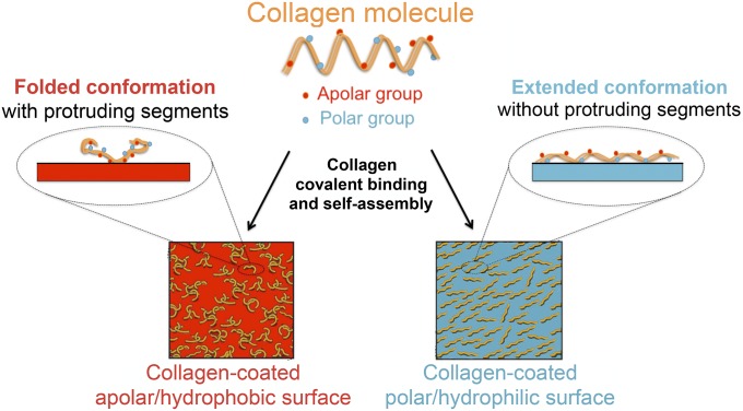
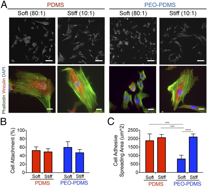
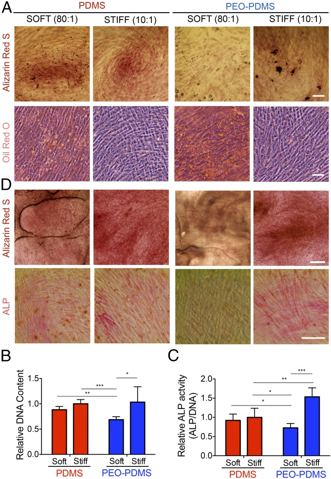
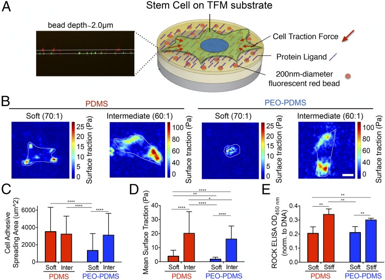
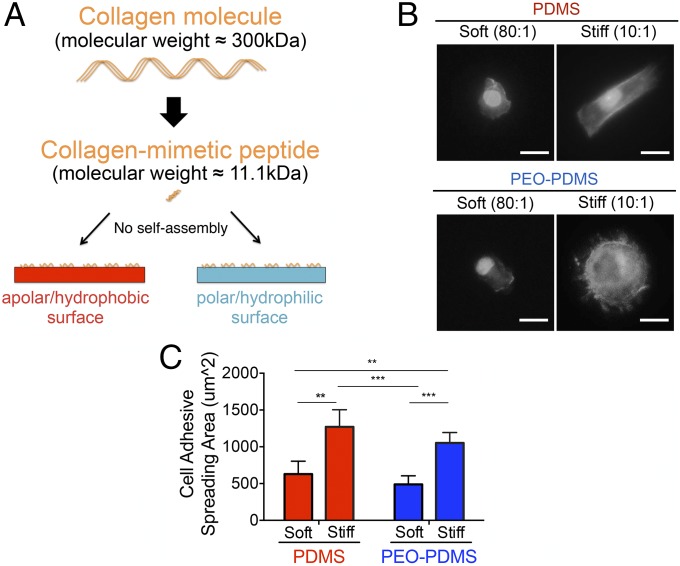
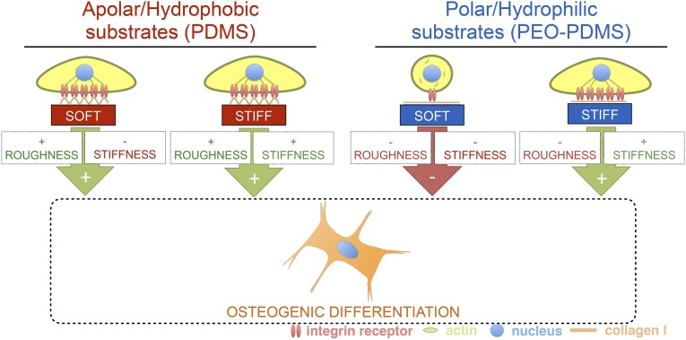
Although mechanisms of cell-material interaction and cellular mechanotransduction are increasingly understood, the mechanical insensitivity of mesenchymal cells to certain soft amorphous biomaterial substrates has remained largely unexplained. We reveal that surface energy-driven supramolecular ligand assembly can regulate mesenchymal stem cell (MSC) sensing of substrate mechanical compliance and subsequent cell fate. Human MSCs were cultured on collagen-coated hydrophobic polydimethylsiloxane (PDMS) and hydrophilic polyethylene-oxide-PDMS (PEO-PDMS) of a range of stiffnesses. Although cell contractility was similarly diminished on soft substrates of both types, cell spreading and osteogenic differentiation occurred only on soft PDMS and not hydrophilic PEO-PDMS (elastic modulus <1 kPa). Substrate surface energy yields distinct ligand topologies with accordingly distinct profiles of recruited transmembrane cell receptors and related focal adhesion signaling. These differences did not differentially regulate Rho-associated kinase activity, but nonetheless regulated both cell spreading and downstream differentiation.

Keywords: PDMS; ligand assembly; mechanobiology; stem cell; surface energy.


Conflict of interest statement

The authors declare no conflict of interest.

Figures

Fig. 1.
Fig. 1.
Schematic of surface energy-driven assembly of collagen I. Collagen I molecules containing polar and apolar amino acid residues covalently bind and self-assemble to exhibit different conformations, topologies, and functionalities when coated on substrates of different surface energy. Adapted from ref. .
Fig. 2.
Fig. 2.
Cell spreading is affected by the surface energy on elastomer substrates of different stiffness. (A) Morphology of hBMSCs on functionalized substrates seeded at 5,000 cells per square centimeter after 24-h culture on PDMS and PEO-PDMS substrates of different stiffness (soft, 0.07–0.10 kPa; stiff, 2.15–2.40 MPa). Cells were immunostained with an antibody against vinculin (red), Alexa-488 phalloidin (green), and DAPI (blue). (Upper row scale bar, 100 μm; Lower row scale bar, 20 μm.) Images of cells on stiff substrates were reported from our previous publication (12). (B) Attachment of hBMSCs on PDMS and PEO-PDMS substrates when seeded at 25,000 cells per square centimeter after 1-h culture (n = 3). Data on stiff substrates were reported from our previous publication (12). (C) hBMSCs spreading area on PDMS and PEO-PDMS when seeded at 5,000 cells per square centimeter after 24-h culture (n = 4; number of cells ≥2,400). Data on stiff substrates were taken from our previous publication (12). Data are represented as mean ± SD. Significance is indicated for P ≤ 0.05 (***P ≤ 0 0.001, ****P ≤ 0.0001).
Fig. 3.
Fig. 3.
Surface energy directs osteogenic stem cell differentiation independently of bulk substrate stiffness. hBMSCs after 7-d culture in mixed-induction medium on PDMS and PEO-PDMS substrates of different stiffness (soft, 0.07–0.10 kPa; stiff, 2.15–2.40 MPa) seeded at 5,000 cells per square centimeter. (A) Staining with Alizarin Red for calcium deposit (Scale bar, 100 μm.) and with Oil Red O for lipid droplets (Scale bar, 200 μm.). (B) Total DNA content. Data on stiff substrates were adapted from our previous studies (12) (n = 4–5). (C) Total ALP activity per DNA normalized by the mean value of the stiff PDMS. Data on stiff substrates were adapted from our previous publication (12) (n = 4–5). (D) Staining of hBMSCs after 14-d culture in basal growth medium on PDMS and PEO-PDMS substrates of different stiffness (soft, 0.07–0.10 kPa; stiff, 2.15–2.40 MPa) seeded at 5,000 cells per square centimeter with Alizarin Red (Scale bar, 500 μm.) and for alkaline phosphatase (ALP) detection with a fast red violet solution. (Scale bar, 100 μm.) Data are represented as mean ± SD. Significance is indicated for P ≤ 0.05 (*P ≤ 0.05, **P ≤ 0.01, ***P ≤ 0.001).
Fig. 4.
Fig. 4.
Traction force microscopy indicates cells spread on soft PDMS without strongly contracting, and is not predominantly mediated by Rho-associated kinase (ROCK) activity. (A) Schematic for PDMS-based TFM platform with embedded 200-nm-diameter fluorescent trackers at a depth of 2.0 μm. (B) Snapshots of traction stress map with color values corresponding to different stress values (see corresponding axis) generated by hBMSCs on PDMS and PEO-PDMS of different stiffness (soft, 0.22–0.35 kPa; intermediate, 5–6 kPa) seeded at 625 cells per square centimeter after 16 h in culture. (Scale bar, 25 μm.) (C) Quantification of the corresponding cell spreading areas with the fluorescent live-cell nucleic acid Syto-13 stain. (n = 56–90). (D) Quantification of the corresponding mean surface traction stresses (see SI Appendix for details about data processing) (n = 56–90). (E) Semiquantification of phosphorylated ROCK by immune-sandwiched enzyme-linked immunosorbent assay when seeded at 5,000 cells per square centimeter after 24-h culture on PDMS and PEO-PDMS substrates of different stiffness (soft, 0.07–0.10 kPa; stiff, 2.15–2.40 MPa) (n = 4–5). Data are represented as mean ± SD. Significance is indicated for P ≤ 0.05 (*P ≤ 0.05, **P ≤ 0.01, ****P ≤ 0.0001).
Fig. 5.
Fig. 5.
Inactivation of collagen self-assembly promotes cell spreading according to PDMS stiffness. (A) Schematic for inactivation of collagen self-assembly by employing a collagen-mimetic (GFOGER) peptide. (B) Morphology of hBMSCs on PDMS and PEO-PDMS substrates of different stiffness (soft, 0.07–0.10 kPa; stiff, 2.15–2.40 MPa) when seeded at 2,500 cells per square centimeter after 24-h staining with Alexa-488 phalloidin and DAPI. (Scale bar, 50 μm.) (C) Quantification of the cell spreading area on the corresponding substrates (n = 4–5; number of cells ≥500). Data are represented as mean ± SD. Significance is indicated for P ≤ 0.05 (**P ≤ 0.01, ***P ≤ 0.001).
Fig. 6.
Fig. 6.
Schematic of the interplay of matrix stiffness and surface energy-driven ligand topography in osteogenic stem cell differentiation.

References

    1. MacQueen L, Sun Y, Simmons CA. Mesenchymal stem cell mechanobiology and emerging experimental platforms. J R Soc Interface. 2013;10:20130179. - PMC - PubMed
    1. Engler AJ, Sen S, Sweeney HL, Discher DE. Matrix elasticity directs stem cell lineage specification. Cell. 2006;126:677–689. - PubMed
    1. Trappmann B, et al. Extracellular-matrix tethering regulates stem-cell fate. Nat Mater. 2012;11:642–649. - PubMed
    1. Wen JH, et al. Interplay of matrix stiffness and protein tethering in stem cell differentiation. Nat Mater. 2014;13:979–987. - PMC - PubMed
    1. Prager-Khoutorsky M, et al. Fibroblast polarization is a matrix-rigidity-dependent process controlled by focal adhesion mechanosensing. Nat Cell Biol. 2011;13:1457–1465. - PubMed

Publication types

LinkOut - more resources