. 2018 May 1;115(18):4678-4683.
doi: 10.1073/pnas.1714938115. Epub 2018 Apr 16.

Deubiquitylation and stabilization of p21 by USP11 is critical for cell-cycle progression and DNA damage responses

Affiliations

Deubiquitylation and stabilization of p21 by USP11 is critical for cell-cycle progression and DNA damage responses

Tanggang Deng et al. Proc Natl Acad Sci U S A. .

Abstract

p21WAF1/CIP1 is a broad-acting cyclin-dependent kinase inhibitor. Its stability is essential for proper cell-cycle progression and cell fate decision. Ubiquitylation by the multiple E3 ubiquitin ligase complexes is the major regulatory mechanism of p21, which induces p21 degradation. However, it is unclear whether ubiquitylated p21 can be recycled. In this study, we report USP11 as a deubiquitylase of p21. In the nucleus, USP11 binds to p21, catalyzes the removal of polyubiquitin chains conjugated onto p21, and stabilizes p21 protein. As a result, USP11 reverses p21 polyubiquitylation and degradation mediated by SCFSKP2, CRL4CDT2, and APC/CCDC20 in a cell-cycle-independent manner. Loss of USP11 causes the destabilization of p21 and induces the G1/S transition in unperturbed cells. Furthermore, p21 accumulation mediated by DNA damage is completely abolished in cells depleted of USP11, which results in abrogation of the G2 checkpoint and induction of apoptosis. Functionally, USP11-mediated stabilization of p21 inhibits cell proliferation and tumorigenesis in vivo. These findings reveal an important mechanism by which p21 can be stabilized by direct deubiquitylation, and they pinpoint a crucial role of the USP11-p21 axis in regulating cell-cycle progression and DNA damage responses.

Keywords: USP11; cell cycle; deubiquitylation; p21; ubiquitin.


Conflict of interest statement

The authors declare no conflict of interest.

Figures
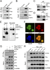
Fig. 1.
Fig. 1.
USP11 interacts with p21. (A and B) A549 cells were transfected with plasmids expressing either Flag-USP11 or Myc-p21. Total cell lysates were subjected to immunoprecipitation with anti-Flag (A) or anti-Myc antibody (B). The immunoprecipitates were then probed with anti-p21, anti-Flag, anti-Myc, or anti-USP11 antibody. (C and D) A549 cell lysates were subjected to immunoprecipitation with control IgG, anti-USP11 (C), or anti-p21 (D) antibody. The immunoprecipitates were then probed with anti-USP11 or anti-p21 antibody. (E) GST, GST-p21, and GST-USP11 produced from bacteria were assessed using Western blotting, and anti-USP11 antibodies were used to immobilize purified GST-USP11 protein onto protein A beads and were then incubated with purified GST or GST-p21. The bound proteins were eluted and analyzed by Western blotting using anti-GST antibodies. (F) The subcellular localization of endogenous USP11 (green) and p21 (red) in A549 cells was visualized using immunofluorescence with anti-USP11 and anti-p21 antibodies. DNA was stained with DAPI, and a merged view of the red and green channels within the same field is shown (merge). (G) A549 cells were transfected with plasmids expressing either Flag-USP11 or the catalytically inactive mutant. Total cell lysates were subjected to immunoprecipitation with anti-Flag antibody. (H) The A549 cells transfected with the indicated Myc-p21 mutants were subjected to immunoprecipitation with anti-Myc antibody.
Fig. 2.
Fig. 2.
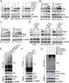
USP11 regulates the protein level of p21. (A) A549, HCT116 WT, and HCT116 p53−/− cells were transfected with the indicated constructs. Total protein was extracted and subjected to Western blotting using anti-Flag, anti-p53, anti-p21, or anti-GAPDH antibody. (B and C) Increasing amounts of USP11 WT or USP11mut were transfected into HCT116 WT (B) and HCT116 p53−/− (C) cells, and total protein was extracted from these cells and subjected to Western blotting using anti-Flag, anti-p53, anti-p21, or anti-GAPDH antibody. (D) A549, HCT116 WT, and HCT116 p53−/− cells were infected with the indicated lentiviral constructs. The resulting cell extracts were analyzed using Western blotting with anti-USP11, anti-p53, anti-p21, or anti-GAPDH antibody. (E and F) HCT116 WT cells either infected with the indicated lentiviral shRNAs (E) or transfected with the indicated constructs (F) were treated with MG132 (20 μM) for 6 h before harvest. p21 was immunoprecipitated with an anti-p21 antibody, and the immunoprecipitates were probed with anti-Ub or anti-p21 antibody. (G) Ubiquitylated Myc-p21 was incubated with GST, GST-tagged USP11 WT, or GST-tagged USP11mut purified from bacteria using glutathione agarose. After coincubation, Myc-p21 was immunoprecipitated using an anti-Myc antibody, and the immunoprecipitates were probed using antibodies against HA and Myc. Recombinant GST, GST-tagged USP11 WT, or GST-tagged USP11mut were analyzed using SDS/PAGE and Coomassie blue staining.
Fig. 3.
Fig. 3.
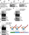
USP11 knockdown abolishes p21 elevation triggered by genotoxic agents. (A and B) HCT116 WT (A) and HCT116 p53−/− (B) cells infected with the indicated lentiviral shRNAs were treated with 5 μM etoposide (Etop) for either 8 or 16 h. Cell lysates were then extracted and subjected to Western blotting. (C) Lysates of HCT116 cells with or without treatment with 5 μM etoposide (Etop) or 0.2 μM doxorubicin (Dox) for 16 h were subjected to immunoprecipitation with control IgG or anti-p21 antibody. The immunoprecipitates were then probed with anti-USP11 or anti-p21 antibody. (D) A proposed working model for p21 regulation by USP11 in response to DNA damage.
Fig. 4.
Fig. 4.
USP11 protects p21 from ubiquitin-mediated degradation. (AC) HCT116 WT cells infected with the indicated shRNAs were transfected with scrambled, SKP2 (A), CDT2 (B), or CDC20 (C) siRNA for 48 h, and then the cell lysates were harvested and analyzed using Western blotting. (DF) HCT116 cells infected with the indicated shRNAs were transfected with scrambled, SKP2 (D), CDT2 (E), and CDC20 (F) siRNA as indicated for 48 h and treated with 20 μM of the proteasome inhibitor MG132 (Sigma) for another 6 h. p21 was immunoprecipitated with an anti-p21 antibody, and the immunoprecipitates were probed with anti-p21 or anti-Ub antibody. (G) A proposed working model that illustrates how USP11 reverses p21 ubiquitination in a cell-cycle–independent manner.
Fig. 5.
Fig. 5.
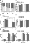
USP11 regulates the G1/S transition and DNA damage-induced G2 checkpoint in a p21-dependent manner. (A) HCT116 WT, HCT116 p53−/−, and HCT116 p21−/− cells infected with the indicated lentiviral shRNAs were stained with propidium iodide and analyzed using flow cytometry. (B) HCT116 WT and HCT116 p21−/− cells transfected with the indicated shRNAs were labeled with BrdU for 60 min before harvesting and then analyzed using flow cytometry. The error bars represent the mean ± SD of three independent experiments. *P < 0.05. (C) HCT116 WT cells infected with the indicated lentiviral shRNAs were transfected with the indicated constructs for 24 h. Cells were stained with propidium iodide and analyzed using flow cytometry. The error bars represent the mean ± SD of three independent experiments. *P < 0.05. (D) HCT116 WT and HCT116 p21−/− cells infected the indicated lentiviral shRNAs were pretreated with 0.2 μM doxorubicin for 2 h, followed by synchronization with nocodazole (100 ng⋅mL−1) for 16 h. The mitotic index was determined using pH3 staining as a marker of mitosis. The error bars represent the mean ± SD of three independent experiments. **P < 0.01. (E and F) HCT116 WT (E) and HCT116 p21−/− (F) cells were infected with the indicated lentiviral shRNAs. Cells were then treated with either 0.2 μM doxorubicin (Dox) or 5 μM etoposide (Etop) for 48 h, followed by flow cytometry analysis of the sub-G1 fraction. The error bars indicate the mean ± SD of three independent experiments. *P < 0.05.

References

    1. Niculescu AB, III, et al. Effects of p21(Cip1/Waf1) at both the G1/S and the G2/M cell cycle transitions: pRb is a critical determinant in blocking DNA replication and in preventing endoreduplication. Mol Cell Biol. 1998;18:629–643. - PMC - PubMed
    1. Brugarolas J, et al. Inhibition of cyclin-dependent kinase 2 by p21 is necessary for retinoblastoma protein-mediated G1 arrest after gamma-irradiation. Proc Natl Acad Sci USA. 1999;96:1002–1007. - PMC - PubMed
    1. Ewen ME, et al. Functional interactions of the retinoblastoma protein with mammalian D-type cyclins. Cell. 1993;73:487–497. - PubMed
    1. Smits VA, et al. p21 inhibits Thr161 phosphorylation of Cdc2 to enforce the G2 DNA damage checkpoint. J Biol Chem. 2000;275:30638–30643. - PubMed
    1. Charrier-Savournin FB, et al. p21-Mediated nuclear retention of cyclin B1-Cdk1 in response to genotoxic stress. Mol Biol Cell. 2004;15:3965–3976. - PMC - PubMed

Publication types

Substances

LinkOut - more resources