. 2018 Apr 17;8(1):6071.
doi: 10.1038/s41598-018-24440-w.

Attenuated DNA damage responses and increased apoptosis characterize human hematopoietic stem cells exposed to irradiation

Affiliations

Attenuated DNA damage responses and increased apoptosis characterize human hematopoietic stem cells exposed to irradiation

Shahar Biechonski et al. Sci Rep. .

Abstract

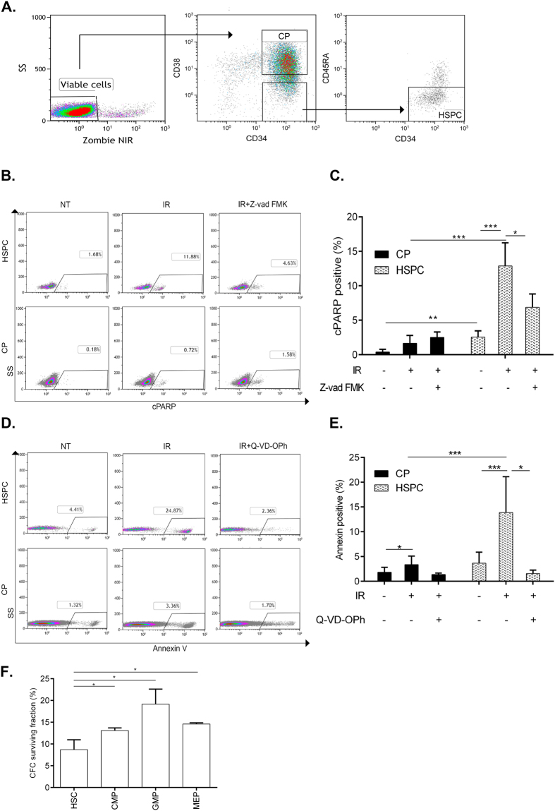
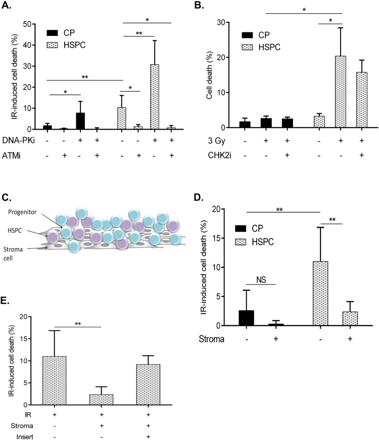
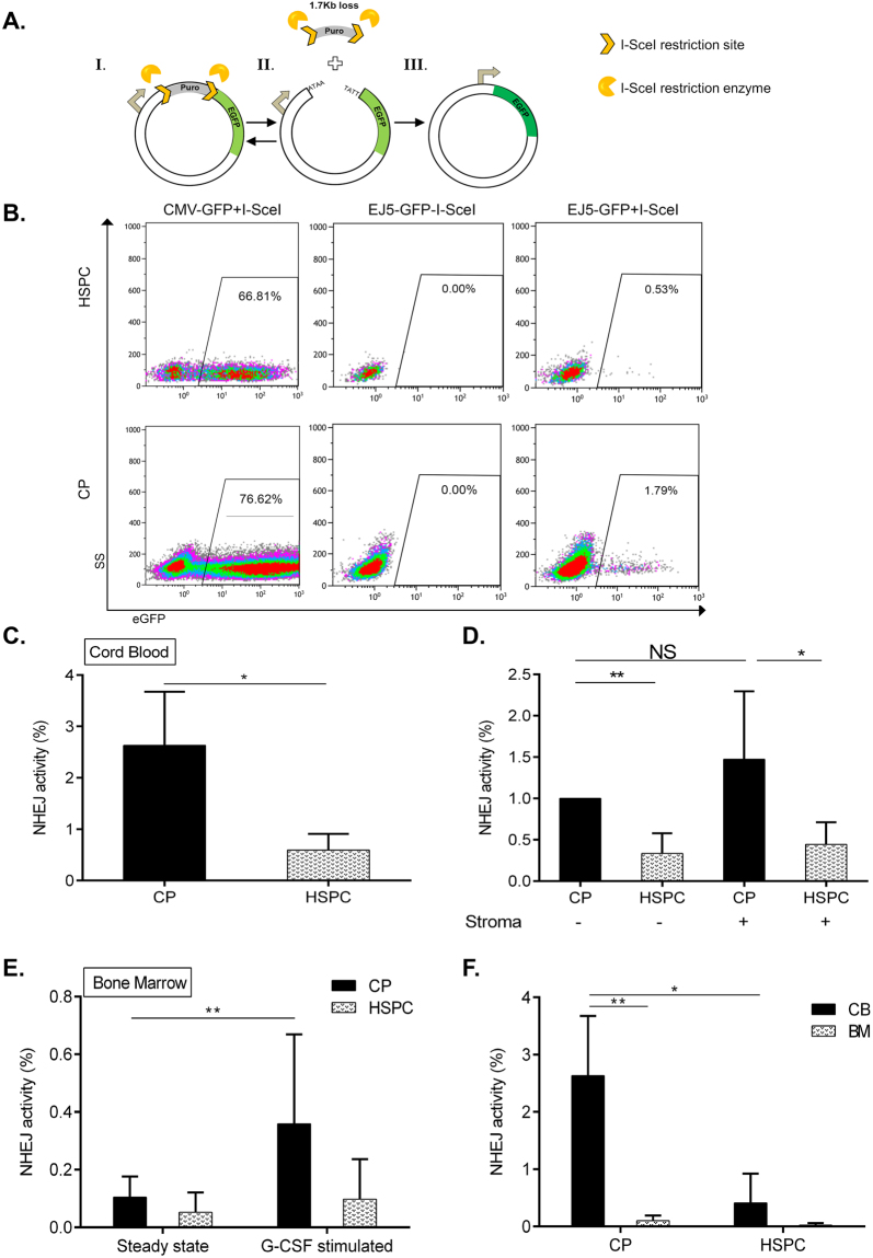
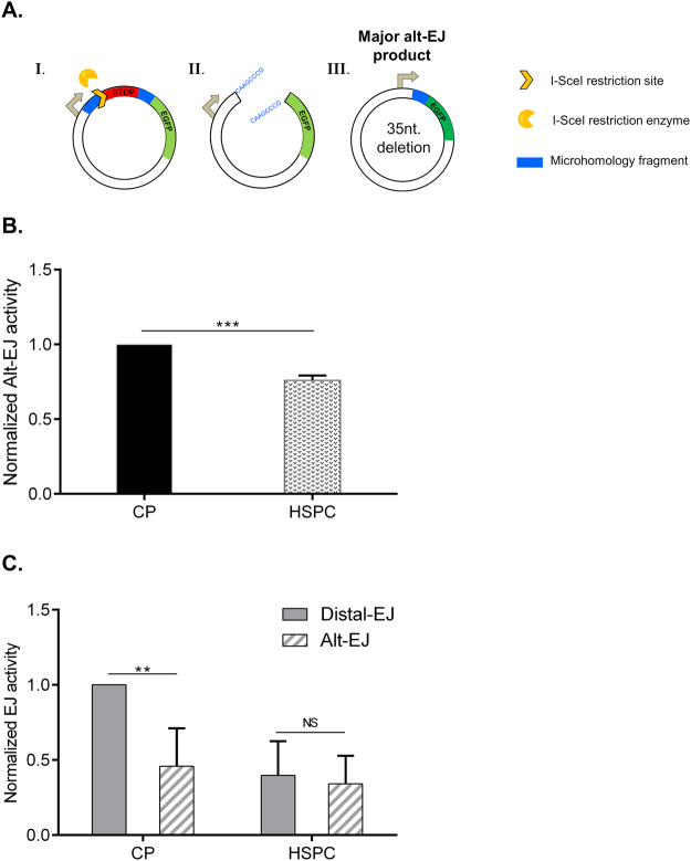
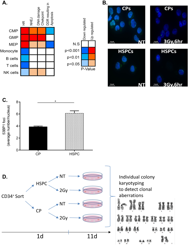
Failure to precisely repair DNA damage in self-renewing Hematopoietic Stem and early Progenitor Cells (HSPCs) can disrupt normal hematopoiesis and promote leukemogenesis. Although HSPCs are widely considered a target of ionizing radiation (IR)-induced hematopoietic injury, definitive data regarding cell death, DNA repair, and genomic stability in these rare quiescent cells are scarce. We found that irradiated HSPCs, but not lineage-committed progenitors (CPs), undergo rapid ATM-dependent apoptosis, which is suppressed upon interaction with bone-marrow stroma cells. Using DNA repair reporters to quantify mutagenic Non-Homologous End Joining (NHEJ) processes, we found that HSPCs exhibit reduced NHEJ activities in comparison with CPs. HSPC-stroma interactions did not affect the NHEJ capacity of HSPCs, emphasizing its cell autonomous regulation. We noted diminished expression of multiple double strand break (DSB) repair transcripts along with more persistent 53BP1 foci in irradiated HSPCs in comparison with CPs, which can account for low NHEJ activity and its distinct control in HSPCs. Finally, we documented clonal chromosomal aberrations in 10% of IR-surviving HSPCs. Taken together, our results revealed potential mechanisms contributing to the inherent susceptibility of human HSPC to the cytotoxic and mutagenic effects of DNA damage.


Conflict of interest statement

The authors declare no competing interests.

Figures

Figure 1
Figure 1
Analysis of IR-induced apoptosis in different fractions of primitive human hematopoietic cells. (A) Flow cytometry gating strategy for HSPCs and CPs. (B,D) Example of FACS data identifying cPARP+ (B) and AnnexinV+ (D) fractions in HSPCs and CPs gated as described in (A) 6 h after 3 Gy IR-treatment. (C,E) CD34+ cells were pre-treated with Z-vad-FMK (100 μM), Q-VD-OPh (20 μM) or DMSO for 1 h, irradiated with 3 Gy and analyzed for cPARP+ or Annexin V+ cells in HSPCs and CPs 6 h later, n ≥ 3. (F) Survival of clonogenic cells from sorted HSPC (CD34+CD38−/lowCD90+45RA), CMP (CD34+CD38+CD45RACD135+), GMP (CD34+CD38+CD45RA+CD135+) and MEP (CD34+CD38+CD45RACD135) fractions following IR with 3 Gy. CFC surviving fraction was calculated by dividing number of colonies counted in the irradiated plates by the number of colonies from the same fraction scored in the non-irradiated plates, n = 3. Represented are mean values ± SD; *p < 0.05, **p < 0.01, ***p < 0.001.
Figure 2
Figure 2
Regulation of IR-induced apoptosis in HSPCs and CPs. (A) Freshly isolated CD34+ cells were pre-treated with ATMi (Ku60019, 10 μM), DNA-PKi (Nu7441, 10 μM) for 1 h, irradiated with 3 Gy and analyzed for cPARP + cells in HSPC and CP fractions 6 h later.n ≥ 4 (B) Freshly isolated CD34+ cells were pre-treated with CHK2i (PV1019, 10 μM) for 1 h, irradiated with 3 Gy and analyzed for Annexin+ cells in HSPCs and CPs 6 h later. n = 3 (C) Schematic presentation of OP9M2 stroma CD34+ co-culture system. (D) Freshly isolated CD34+ cells were irradiated (3 Gy) or not and immediately plated on pre-established confluent layers of OP9M2 stromal cells for 6 hrs followed by Annexin+ cells analysis in HSPC and CP gates. IR-induced cell death was calculated to reduce the variability between CD34+ batches by subtracting the fraction of c-PARP+ or AnnexinV+ cells scored in the untreated sample from the same fraction in the IR sample n = 7 (E) CD34+ cells were plated on OP9M2 stroma as described in (D) in the presences or absence of pore (0.4 µm) membrane insert, n ≥ 3. Represented are mean values ± SD, *p < 0.05, **p < 0.01.
Figure 3
Figure 3
Analysis of DSB repair via NHEJ in CPs and HSPCs using a reporter system. (A) Schematic presentation of DSB repair substrate EJ5-GFP (I.) and I-SceI endonuclease-mediated cleavage (II.), triggering NHEJ processes which restore the EGFP expression cassette (III.). (B) Representative flow charts of NHEJ activity and nucleofection efficiency in HSPCs and CPs. Freshly isolated CD34+ cells from cord blood were nucleofected with plasmid CMV-GFP (positive control for EGFP expression) or EJ5-GFP reporter with or without I-SceI endonuclease expression plasmid and cultured for 24 h. Then, the proportion of viable EGFP + cells in HSPC- and CP- fractions gated as described in Fig. 1A was analyzed by flow cytometry. (C) NHEJ capacity of CP- and HSPC- enriched fractions isolated from cord blood (n = 3). (D) Nucleofected CD34+ cells from cord blood were plated in wells containing medium only or stroma layers for 24 hrs, followed by NHEJ capacity assessment in CP- and HSPC- enriched fractions. (E) Human bone marrow samples obtained from G-CSF treated (10 ug/kg for 5 days prior to harvesting) (n = 2) or steady-state control donors (n = 4) were nucleofected and processed for NHEJ activity assessment as described in Fig. 1B. NHEJ frequencies were individually normalized to the nucleofection efficiencies in each subpopulation and experiment. (F) Comparison of NHEJ frequencies of HSPCs and CPs derived from cord blood versus untreated bone marrow based on measurements presented in (C) and (E). Represented are mean values ± SD; *p value < 0.05, **p value < 0.01, NS, non-significant.
Figure 4
Figure 4
Alt-EJ activity and its regulation in CPs and HSPCs. (A) Schematic presentation of DSB repair reporter EJ2-GFP (I.), repair intermediate induced by I-SceI endonuclease (II.) and EGFP + product obtained via Alt-EJ (III.). (B) Freshly isolated CD34+ cells were nucleofected with EJ2-GFP reporter together with I-SceI expression plasmid and cultured for 24 h. Then, the fraction of EGFP + cells in viable CPs and HSPCs cell fractions gated as described in Fig. 1A was analyzed by flow cytometry, n = 3. (C) Comparison of NHEJ and Alt-EJ capacity in CPs and HSPCs based on measurements presented in Figs. 3C and 4B. The proportion of Alt-EJ events (measured by EJ2-GFP assay) were calculated by dividing EJ2-GFP readout on the EJ5-GFP readout for the same cell fraction. Represented are mean values ± SD, **p value < 0.01, ***p < 0.001, NS; non-significant.
Figure 5
Figure 5
DNA repair and DDR gene expression, DSB marker and genome stability analyses. (A) left panel: relative expression of transcripts related to DNA repair, DDR checkpoint and apoptosis pathways in CPs (CMP, GMP and MEP) and mature blood-derived cells (B, T and NK) in comparison to HSPCs colored accordingly to the significance of the changes. right panel: color scheme description with blue colors for decreased expression and red colors indicative of increased expression relative to HSPCs along with the corresponding p values. (B) CD34+ cells were sorted into HSPC and CP fractions. Representative immunofluorescence staining visualized for 53BP1 (green) and DNA (blue) in HSPCs and CPs 6 hr after 3 Gy IR. (C) Average number of 53BP1 foci per cell in CPs and HSPCs. n = 3 experiments; Represented are mean values ± SD; *p < 0.05. At least 100 nuclei were analyzed in each experiment. (D) Workflow for the karyotype analysis of irradiated HSPCs and CPs. Freshly isolated CD34+ cells were sorted for HSPC and CP fractions, exposed or not to IR (2 Gy) and then plated at low density in methylcellulose to allow colony formation. Individual colonies were isolated for metaphase preparation and karyotype analysis (Table 1, and Supplementary Table 1).

References

    1. Welch JS, et al. The origin and evolution of mutations in acute myeloid leukemia. Cell. 2012;150:264–278. doi: 10.1016/j.cell.2012.06.023. - DOI - PMC - PubMed
    1. Jan M, et al. Clonal evolution of preleukemic hematopoietic stem cells precedes human acute myeloid leukemia. Science translational medicine. 2012;4:149ra118. doi: 10.1126/scitranslmed.3004315. - DOI - PMC - PubMed
    1. Xie M, et al. Age-related mutations associated with clonal hematopoietic expansion and malignancies. Nat Med. 2014;20:1472–1478. doi: 10.1038/nm.3733. - DOI - PMC - PubMed
    1. Shlush LI, et al. Identification of pre-leukaemic haematopoietic stem cells in acute leukaemia. Nature. 2014 - PMC - PubMed
    1. Genovese G, et al. Clonal Hematopoiesis and Blood-Cancer Risk Inferred from Blood DNA Sequence. New England Journal of Medicine. 2014;371:141126135947008–141126135947008. doi: 10.1056/NEJMoa1409405. - DOI - PMC - PubMed

MeSH terms