Review
. 2018 Jun;19(6):323-337.
doi: 10.1038/s41583-018-0001-8.

Intrinsic mechanisms of neuronal axon regeneration

Affiliations
Review

Intrinsic mechanisms of neuronal axon regeneration

Marcus Mahar et al. Nat Rev Neurosci. 2018 Jun.

Abstract

Permanent disabilities following CNS injuries result from the failure of injured axons to regenerate and rebuild functional connections with their original targets. By contrast, injury to peripheral nerves is followed by robust regeneration, which can lead to recovery of sensory and motor functions. This regenerative response requires the induction of widespread transcriptional and epigenetic changes in injured neurons. Considerable progress has been made in recent years in understanding how peripheral axon injury elicits these widespread changes through the coordinated actions of transcription factors, epigenetic modifiers and, to a lesser extent, microRNAs. Although many questions remain about the interplay between these mechanisms, these new findings provide important insights into the pivotal role of coordinated gene expression and chromatin remodelling in the neuronal response to injury.


Figures
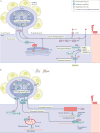
Fig. 1
Fig. 1. Signalling events following peripheral axon injury
a | Early injury signalling events. Following injury, calcium rapidly flows into the damaged axon and is actively propagated to the cell soma by voltage-gated calcium channels (VGCCs) and the release of calcium by internal stores in the endoplasmic reticulum (ER). The release of stored calcium also triggers cellular stress pathways in the axon, resulting in the splicing of X-box-binding protein 1 (XBP1) and its translocation to the nucleus. The increase in calcium activates pro-regenerative pathways at the site of injury and in the cell soma. For example, near the injury site, calcium activates cAMP, which in turn activates protein kinase A (PKA). PKA then activates dual leucine zipper-bearing kinase (DLK), a key mediator of later regenerative events. Similarly, calcium at the injury site facilitates both the local translation of signal transducer and activator of transcription 3 (STAT3), importin-β and filamin A, and the association of extracellular-signal-related kinase (ERK) with importin-β, which allows them to be retrogradely transported. The backpropagation of calcium to the cell soma promotes the translocation of serine/ threonine-protein kinase D1 (PRKD1) to the nucleus, which promotes histone deacetylase 5 (HDAC5) nuclear export. The calcium backpropagation also facilitates the expression of the methylcytosine dioxygenase TET3, which promotes post-injury demethylation of DNA. In addition to the actions of calcium, other pathways are triggered by the injury: in the axon, the transcription factor STAT3 is phosphorylated, probably through the activity of cytokines, and is retrogradely transported to the nucleus to activate regenerative programmes. b | Late injury signalling events. In the axon, DLK enables the retrograde transport of several injury signalling proteins, including JUN N-terminal kinase (JNK), JNK-interacting protein 3 (JIP3) and DLK itself, to the nucleus,,. This process is also facilitated by an increase in tyrosinated tubulin near the injury site. Phosphorylated STAT3 can localize either to mitochondria (when phosphorylated at S727) to increase ATP synthesis or to the nucleus (when phosphorylated at Y705) to influence transcription of pro-regenerative genes. ERK, which is protected by vimentin during its retrograde transport, causes the histone acetyltransferase KAT2B to translocate into the nucleus,. Conversely, HDAC5 and HDAC3 are exported from the nucleus in response to PRKD1 nuclear translocation and HDAC5 is transported to the growth cone, where it interacts with filamin A and PRKD1 to deacetylate microtubules,. The changes in subcellular location of these histone modifiers, along with the calcium-dependent increase in TET3 (REF.), cause a transition from repressive DNA and histone methylation (Me) into permissive histone acetylation (Ac). This transition likely results in a relaxation of the DNA around pro-regenerative genes, allowing binding by pro-regenerative transcription factors and expression of pro-regenerative genes,,. Additional signalling may also be provided by surrounding satellite glial cells, which respond to axon injury by downregulating the ATP-dependent inwardly rectifying potassium channel Kir4.1 (KCNJ10) and upregulating glial fibrillary acidic protein (GFAP). The molecular mechanisms by which satellite glial cells can sense a distant axon injury remain largely unknown (indicated by the question mark). P, phosphorylation.
Fig. 2
Fig. 2. Model of epigenetic activation of pro-regenerative genes in response to axon injury
a | When dorsal root ganglion neurons have formed synapses and are electrically active, pro-regenerative gene bodies, promoters and enhancers are maintained in a transcriptionally repressed state through DNA methylation by DNA (cytosine-5)-methyltransferases (DNMTs), histone methylation by yet unknown histone methyltransferases and deacetylation by histone deacetylases (HDACs),,. b | Following injury, the nuclear export of HDACs and import of the histone acetyltransferase (HAT) KAT2B causes an increase in acetylation of histone H3K9 (H3K9Ac), whereas the upregulation of the methylcytosine dioxygenase TET3 causes the conversion of repressive 5-methylcytosine (5mC) into 5-hydroxymethylcytosine (5hmC). Similarly, CREB-binding protein (CBP) and p300 HAT, HATs that frequently bind to both enhancers and promoters, associate with cellular tumour antigen p53 (TP53) to increase H3K9Ac in pro-regenerative gene promoters,. The turnover of transcriptionally repressive marks into activating marks likely causes a relaxation in the chromatin, which should allow pro-regenerative transcription factors — such as Myc proto-oncogene protein (MYC), cAMP-dependent transcription factor ATF3, transcription factor AP-1 (JUN) and signal transducer and activator of transcription 3 (STAT3) — to bind regulatory elements and influence the transcription of downstream pro-regenerative genes. H3K9Me, methylation of histone HeK9; P, phosphorylation; RNAPII, RNA polymerase II.

References

    1. Liu K, Tedeschi A, Park KK, He Z. Neuronal intrinsic mechanisms of axon regeneration. Annu. Rev. Neurosci. 2011;34:131–152. - PubMed
    1. Di Giovanni S. Molecular targets for axon regeneration: focus on the intrinsic pathways. Expert Opin. Ther. Targets. 2009;13:1387–1398. - PubMed
    1. Schwab ME, Strittmatter SM. Nogo limits neural plasticity and recovery from injury. Curr. Opin. Neurobiol. 2014;27:53–60. - PMC - PubMed
    1. Tedeschi A, Bradke F. Spatial and temporal arrangement of neuronal intrinsic and extrinsic mechanisms controlling axon regeneration. Curr. Opin. Neurobiol. 2017;42:118–127. - PubMed
    1. Kaplan A, Bueno M, Hua L, Fournier AE. Maximizing functional axon repair in the injured central nervous system: lessons from neuronal development. Dev. Dyn. 2017;247:18–23. - PubMed

Publication types