Review
. 2018 Apr 3:9:633.
doi: 10.3389/fimmu.2018.00633. eCollection 2018.

Chemotactic Cues for NOTCH1-Dependent Leukemia

Affiliations
Review

Chemotactic Cues for NOTCH1-Dependent Leukemia

Erich Piovan et al. Front Immunol. .

Abstract

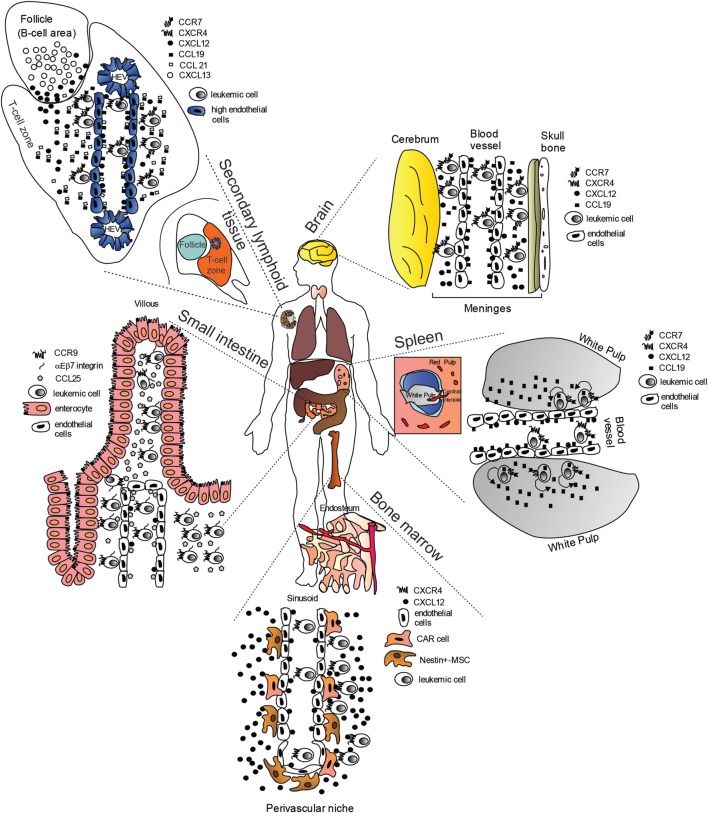
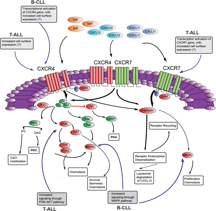
The NOTCH signaling pathway is a conserved signaling cascade that regulates many aspects of development and homeostasis in multiple organ systems. Aberrant activity of this signaling pathway is linked to the initiation and progression of several hematological malignancies, exemplified by T-cell acute lymphoblastic leukemia (T-ALL). Interestingly, frequent non-mutational activation of NOTCH1 signaling has recently been demonstrated in B-cell chronic lymphocytic leukemia (B-CLL), significantly extending the pathogenic significance of this pathway in B-CLL. Leukemia patients often present with high-blood cell counts, diffuse disease with infiltration of the bone marrow, secondary lymphoid organs, and diffusion to the central nervous system (CNS). Chemokines are chemotactic cytokines that regulate migration of cells between tissues and the positioning and interactions of cells within tissue. Homeostatic chemokines and their receptors have been implicated in regulating organ-specific infiltration, but may also directly and indirectly modulate tumor growth. Recently, oncogenic NOTCH1 has been shown to regulate infiltration of leukemic cells into the CNS hijacking the CC-chemokine ligand 19/CC-chemokine receptor 7 chemokine axis. In addition, a crucial role for the homing receptor axis CXC-chemokine ligand 12/CXC-chemokine receptor 4 has been demonstrated in leukemia maintenance and progression. Moreover, the CCL25/CCR9 axis has been implicated in the homing of leukemic cells into the gut, particularly in the presence of phosphatase and tensin homolog tumor suppressor loss. In this review, we summarize the latest developments regarding the role of NOTCH signaling in regulating the chemotactic microenvironmental cues involved in the generation and progression of T-ALL and compare these findings to B-CLL.

Keywords: CXC-chemokine receptor 4; CXCR7; NOTCH; T-cell acute lymphoblastic leukemia; chemokines; infiltration; stromal-derived factor-1.


Figures

Figure 1
Figure 1
“Cellular highways” hijacked by leukemic cells implicated in T-cell acute lymphoblastic leukemia dissemination (many of the findings may also apply to B-cell chronic lymphocytic leukemia). Under physiological conditions, homeostatic chemokines control cellular migration by directing cells expressing specific chemokine receptors to appropriate locations expressing their cognate chemokine ligands. These cellular highways are also used by leukemic cells. In the brain, CC-chemokine ligand 19 (CCL19) and CXC-chemokine ligand 12 (CXCL12) recruit CC-chemokine receptor 7 (CCR7)- and CXC-chemokine receptor 4 (CXCR4)-expressing leukemic cells from blood vessels. In the spleen, CCL19 recruits CCR7-expressing leukemic cells from blood vessels possibly in combination with CXCL12. Migrated leukemic cells may then activate an autocrine/paracrine secretion of CCL19. CCR7-expressing leukemic cells together with CD62L (not shown) and CXCR4, gain access to secondary lymphoid organs such as lymph nodes (shown) via interactions with CCL19, CCL21, peripheral lymph node vascular addressin (not shown) and CXCL12 presented on high-endothelial venules (HEV). Here, leukemic cells are retained, proliferate, and completely substitute the normal tissue architecture. In the bone marrow (BM), CXCR4-expressing leukemic cells are probably initially recruited to the perivascular niche expressing high levels CXCL12, where a leukemic niche is established. Inhibitors of the CXCL12/CXCR4 interaction release leukemic cells from their BM niche, and allow these cells to enter the blood stream. In the small intestine, CCR9-expressing leukemia cells (together with αEβ7 integrin) are recruited by CCL25, where the presence of phosphoinositide-3 kinase-AKT pathway activation contributes to confer a proliferative advantage to leukemic cells in an otherwise non-supportive microenvironment.
Figure 2
Figure 2
Schematic diagram of putative CXCR4–CXCR7 crosstalk affecting signaling pathways. The influence of NOTCH signaling on the main aspects of this signaling axis is shown in gray boxes [differences between T-cell acute lymphoblastic leukemia (T-ALL) and B-cell chronic lymphocytic leukemia (B-CLL) is presented]. CXCL12 employs two distinct receptors, CXCR4 and CXCR7 which can form homodimers or heterodimers. Additionally, CXCR4 and CXCR7 can act as receptors for macrophage migration inhibitory factor (MIF), while CXCR7 can also bind to CXCL11. Commonly, stimulation of CXCR4 leads to G-protein-coupled chemokine receptors (GPCR) signaling through phosphoinositide-3 kinase (PI3K)/AKT, PLC/IP3, MAPK pathways, and mobilization of Ca2+ from intracellular sources. CXCR4/CXCR7 heterodimerization attenuates GPCR signaling, promoting β-arrestin mediated signaling. Activation of CXCR7 triggers β-arrestin mediated signaling. Internalization of the receptors CXCR4 and CXCR7, and subsequent recycling to the cell surface, is also mediated by β-arrestin. Binding of CXCL12 to CXCR7 promotes internalization and scavenging (lysosomal degradation) of CXCL12. AC, adenylyl cyclase; cAMP, cyclic adenosyl monophosphate; PKA, protein kinase A; PLC, phospholipase C; GRK, GPCR kinase; PI3K, phosphatidylinositide 3-kinase; Gα/Gβ/Gγ, heterotrimeric G-protein consisting of subunits α, β, and γ; PIP2, phosphatidylinositol 4,5-bisphosphate; IP3, inositol 1,4,5-bisphosphate; AKT, protein kinase B; MAPK, mitogen-activated protein kinase; FAK, focal adhesion kinase; Pyk-2, proline rich kinase-2; DAG, diacylglycerol; PKC, protein kinase C. “?”, not known; black, pathway activation; red, pathway repression.

References

    1. Artavanis-Tsakonas S, Rand MD, Lake RJ. Notch signaling: cell fate control and signal integration in development. Science (1999) 284(5415):770–6.10.1126/science.284.5415.770 - DOI - PubMed
    1. D’Souza B, Miyamoto A, Weinmaster G. The many facets of Notch ligands. Oncogene (2008) 27(38):5148–67.10.1038/onc.2008.229 - DOI - PMC - PubMed
    1. Andersson ER, Sandberg R, Lendahl U. Notch signaling: simplicity in design, versatility in function. Development (2011) 138(17):3593–612.10.1242/dev.063610 - DOI - PubMed
    1. Koch U, Radtke F. Notch and cancer: a double-edged sword. Cell Mol Life Sci (2007) 64(21):2746–62.10.1007/s00018-007-7164-1 - DOI - PMC - PubMed
    1. Radtke F, Raj K. The role of Notch in tumorigenesis: oncogene or tumour suppressor? Nat Rev Cancer (2003) 3(10):756–67.10.1038/nrc1186 - DOI - PubMed