. 2018 Apr 13:6:e4593.
doi: 10.7717/peerj.4593. eCollection 2018.

Evolution of dopamine receptors: phylogenetic evidence suggests a later origin of the DRD2l and DRD4rs dopamine receptor gene lineages

Affiliations

Evolution of dopamine receptors: phylogenetic evidence suggests a later origin of the DRD2l and DRD4rs dopamine receptor gene lineages

Juan C Opazo et al. PeerJ. .

Abstract

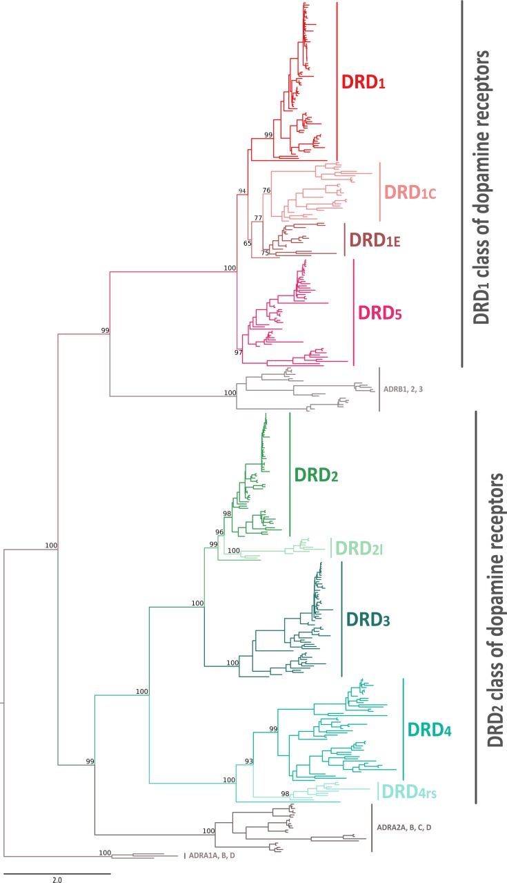
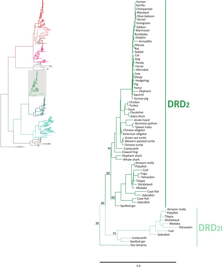
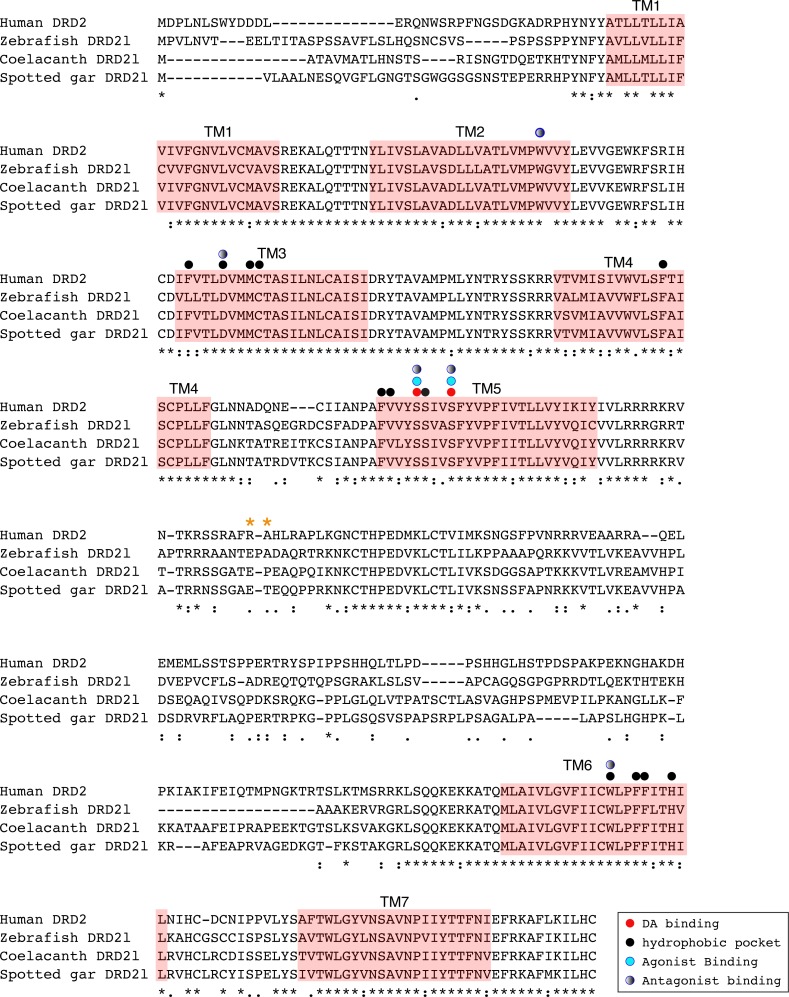
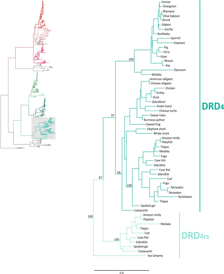
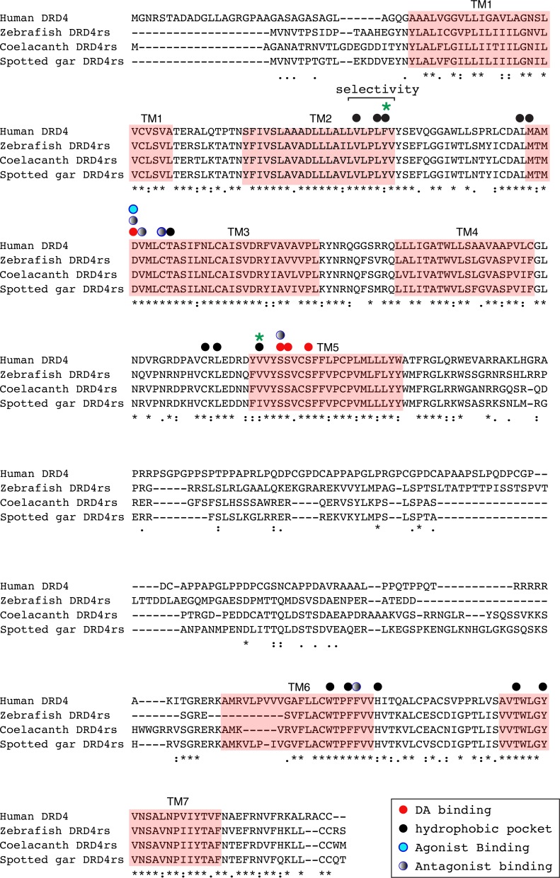
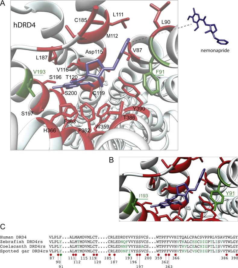
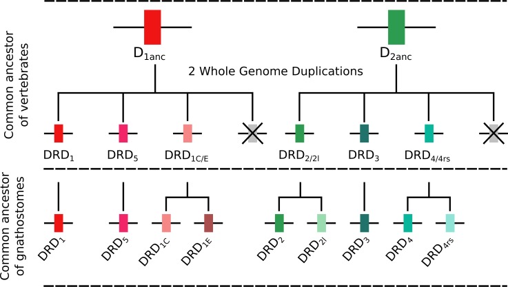
Dopamine receptors are integral membrane proteins whose endogenous ligand is dopamine. They play a fundamental role in the central nervous system and dysfunction of dopaminergic neurotransmission is responsible for the generation of a variety of neuropsychiatric disorders. From an evolutionary standpoint, phylogenetic relationships among the DRD1 class of dopamine receptors are still a matter of debate as in the literature different tree topologies have been proposed. In contrast, phylogenetic relationships among the DRD 2 group of receptors are well understood. Understanding the time of origin of the different dopamine receptors is also an issue that needs further study, especially for the genes that have restricted phyletic distributions (e.g., DRD2l and DRD4rs). Thus, the goal of this study was to investigate the evolution of dopamine receptors, with emphasis on shedding light on the phylogenetic relationships among the D1 class of dopamine receptors and the time of origin of the DRD2l and DRD4rs gene lineages. Our results recovered the monophyly of the two groups of dopamine receptors. Within the DRD1 group the monophyly of each paralog was recovered with strong support, and phylogenetic relationships among them were well resolved. Within the DRD1 class of dopamine receptors we recovered the sister group relationship between the DRD1C and DRD1E, and this clade was recovered sister to a cyclostome sequence. The DRD1 clade was recovered sister to the aforementioned clade, and the group containing DRD5 receptors was sister to all other DRD1 paralogs. In agreement with the literature, among the DRD2 class of receptors, DRD2 was recovered sister to DRD3, whereas DRD4 was sister to the DRD2/DRD3 clade. According to our phylogenetic tree, the DRD2l and DRD4rs gene lineages would have originated in the ancestor of gnathostomes between 615 and 473 mya. Conservation of sequences required for dopaminergic neurotransmission and small changes in regulatory regions suggest a functional refinement of the dopaminergic pathways along evolution.

Keywords: Dopamine receptors; Gene family evolution; Neuroscience; Whole genome duplications.


Conflict of interest statement

The authors declare there are no competing interests.

Figures

Figure 1
Figure 1. Maximum likelihood tree depicting evolutionary relationships among dopamine receptors in vertebrates.
Numbers on the nodes correspond to maximum likelihood ultrafast bootstrap support values. Human ADRA1A, ADRA1B, and ADRA1D sequences were used as outgroups.
Figure 2
Figure 2. Patterns of conserved synteny in the chromosomal regions that harbor the DRD1 class of dopamine receptors.
(A) Chromosomal region that harbors the DRD1 gene; (B) Chromosomal region that harbors the DRD1C gene; (C) Chromosomal region that harbors the DRD1E gene; (D) Chromosomal region that harbors the DRD5 gene. Asterisks denote that the orientation of the genomic piece is from 3′ to 5′, gray lines represent intervening genes that do not contribute to conserved synteny whereas dashed lines represent genes that are not present.
Figure 3
Figure 3. Patterns of conserved synteny in the chromosomal regions that harbor the DRD2 class of dopamine receptors.
(A) Chromosomal region that harbors DRD2 gene; (B) Chromosomal region that harbors DRD21 gene; (C) Chromosomal region that harbors DRD3 gene; (D) Chromosomal region that harbors DRD4 gene; (E) Chromosomal region that harbors DRD4rs gene. Asterisks denote that the orientation of the genomic piece is from 3′ to 5′, gray lines represent intervening genes that do not contribute to conserved synteny whereas dashed lines represent genes that are not present.
Figure 4
Figure 4. Maximum likelihood trees depicting evolutionary relationships among DRD2 and DRD2l dopamine receptors in vertebrates.
Numbers on the nodes correspond to maximum likelihood ultrafast bootstrap support values. This tree topology does not represent novel phylogenetic analyses; they are the DRD2/DRD2l clade that was recovered from Fig. 1.
Figure 5
Figure 5. Phyletic distribution of dopamine receptor genes in vertebrates.
The cyclostome check between the DRD1C and DRDE indicates that the duplication event that gave rise to these genes occurred after the divergence between cyclostomes and gnathostomes. Therefore, cyclostomes retained the ancestral condition of a single gene copy. A similar situation applies to DRD2/DRD2l and to DRD4/DRD4rs.
Figure 6
Figure 6. Alignment of the human dopamine receptor 2 (DRD2) with zebrafish (Danio rerio), coelacanth (Latimeria chalumnae) and spotted gar (Lepisosteus oculatus) dopamine receptor 2l (DRD2l).
Shaded regions denote transmembrane domains according to UniProt. Dopamine binding sites, agonist and antagonist binding sites were predicted with theoretical and computational techniques (Yashar et al., 2004) and experimental evidence (Shi & Javitch, 2002) . Amino acids in the third intracellular loop conferring G protein subunit Gαi specificity (Senogles et al., 2004) are indicated by orange asterisks.
Figure 7
Figure 7. Maximum likelihood trees depicting evolutionary relationships among DRD4 and DRD4rs dopamine receptors in vertebrates.
Numbers on the nodes correspond to maximum likelihood ultrafast bootstrap support values. This tree topology does not represent novel phylogenetic analyses; they are the DRD4/DRD4rs clade that was recovered from Fig. 1.
Figure 8
Figure 8. Alignment of the human dopamine receptor 4 (DRD4) with zebrafish (Danio rerio), coelacanth (Latimeria chalumnae) and spotted gar (Lepisosteus oculatus) dopamine receptor 4rs (DRD4rs).
Shaded regions denote transmembrane domains according to UniProt. Dopamine binding sites (red dots) were determined by site directed mutagenesis (Cummings et al., 2010) and homology to DRD2. Antagonist binding sites and hydrophobic pocket-including selectivity region-were obtained from mutagenesis studies (Cummings et al., 2010) and from the crystal structure of the receptor coupled to the antagonist nemonapride (Wang et al., 2017). Non-conserved amino acids in the nemonapride binding pocket are labeled with green asterisks. Binding sites for the selective agonist UCSF924 are also shown (light blue dot).
Figure 9
Figure 9. Structural details of human DRD4 binding site to the antipsychotic drug nemonapride (in blue) based on the molecular file PDB ID: 5WIV (Wang et al., 2017).
(B) Conserved amino acids within 4 Å of the drug molecule are shown with functional groups (in red). Non-conserved amino acids (in green) were changed (inset A) to the residue present in the fish species: F91Y and V193I. This mutagenesis was simulated choosing the rotamer (orientation of the side chain) with the highest probability (Y rotamer: 72.6%; I rotamer: 79% probability) see methods for additional details. (C) Partial alignment of the human dopamine receptor 4 (DRD4) with zebrafish (Danio rerio), coelacanth (Latimeria chalumnae) and spotted gar (Lepisosteus oculatus) dopamine receptor 4rs (DRD4rs) showing the numbers corresponding to the human DRD4 sequence (NP_000788). Conserved and non-conserved aminoacids shown in (B) are indicated with red and green dots respectively. Non-conserved aminoacids within the region are also shown in green fonts.
Figure 10
Figure 10. An evolutionary hypothesis regarding the origin of dopamine receptor genes in vertebrates.
The vertebrate ancestor possessed two dopamine receptors, one of each class. However, after the two rounds of whole genome duplications (WGD) that occurred in the ancestor of the group each ancestral gene (DRD1anc and DRD2anc) gave rise to four genes. In the case of the DRD1 class of receptors three out of the four genes were retained in the genome of the vertebrate ancestor. In the gnathostome ancestor, the DRD1C∕E gene underwent a duplication event that gave rise to the actual DRD1C and DRD1E genes. Thus, the gnathostome ancestor had a repertoire of four DRD1 genes: DRD1, DRD5, DRD1C and DRD1E. In the case of the DRD2 group of receptors, the vertebrate WGDs originated four genes, three of which were maintained in the genome of extant species (DRD2∕2l, DRD3 and DRD4∕4rs). In the ancestor of gnathostomes, the DRD2∕2l gene underwent a duplication event that gave rise to an extra copy of the gene. Similarly, the DRD4∕4rs gene also underwent a duplication event that gave rise to an extra copy of the gene. Thus, the ancestor of gnathostome vertebrates possessed a repertoire of five DRD2 genes: DRD2, DRD2l, DRD3, DRD4 and DRD4rs.

References

    1. Altschul SF, Gish W, Miller W, Myers EW, Lipman DJ. Basic local alignment search tool. Journal of Molecular Biology. 1990;215:403–410. doi: 10.1016/S0022-2836(05)80360-2. - DOI - PubMed
    1. Araya R, Andino-Pavlovsky V, Yuste R, Etchenique R. Two-photon optical interrogation of individual dendritic spines with caged dopamine. ACS Chemical Neuroscience. 2013;4:1163–1167. doi: 10.1021/cn4000692. - DOI - PMC - PubMed
    1. Ashok AH, Marques TR, Jauhar S, Nour MM, Goodwin GM, Young AH, Howes OD. The dopamine hypothesis of bipolar affective disorder: the state of the art and implications for treatment. Molecular Psychiatry. 2017;22:666–679. doi: 10.1038/mp.2017.16. - DOI - PMC - PubMed
    1. Beaulieu J-M, Gainetdinov RR. The physiology, signaling, and pharmacology of dopamine receptors. Pharmacological Reviews. 2011;63:182–217. doi: 10.1124/pr.110.002642. - DOI - PubMed
    1. Boehmier W, Obrecht-Pflumio S, Canfield V, Thisse C, Thisse B, Levenson R. Evolution and expression of D2 and D3 dopamine receptor genes in zebrafish. Developmental Dynamics. 2004;230:481–493. doi: 10.1002/dvdy.20075. - DOI - PubMed

LinkOut - more resources