Review
. 2018 Apr 3:6:34.
doi: 10.3389/fcell.2018.00034. eCollection 2018.

Cell Chirality Drives Left-Right Asymmetric Morphogenesis

Affiliations
Review

Cell Chirality Drives Left-Right Asymmetric Morphogenesis

Mikiko Inaki et al. Front Cell Dev Biol. .

Abstract

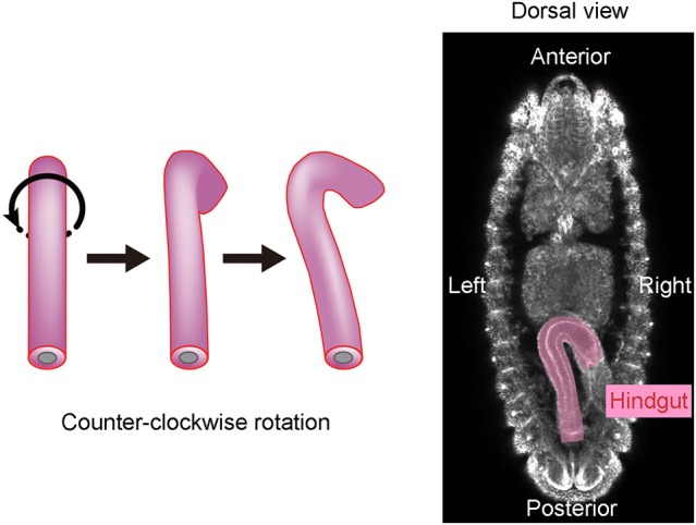
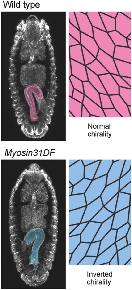
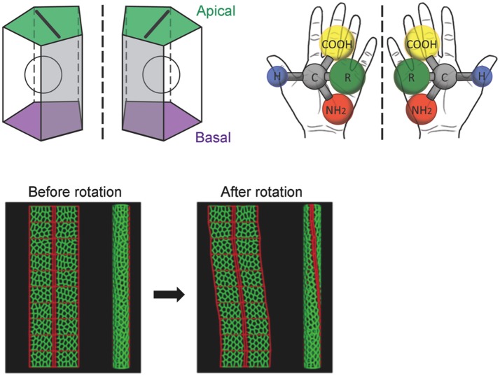
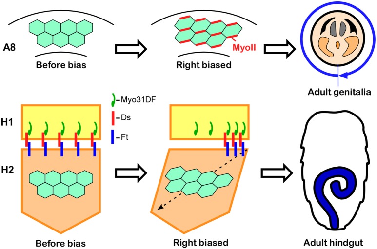
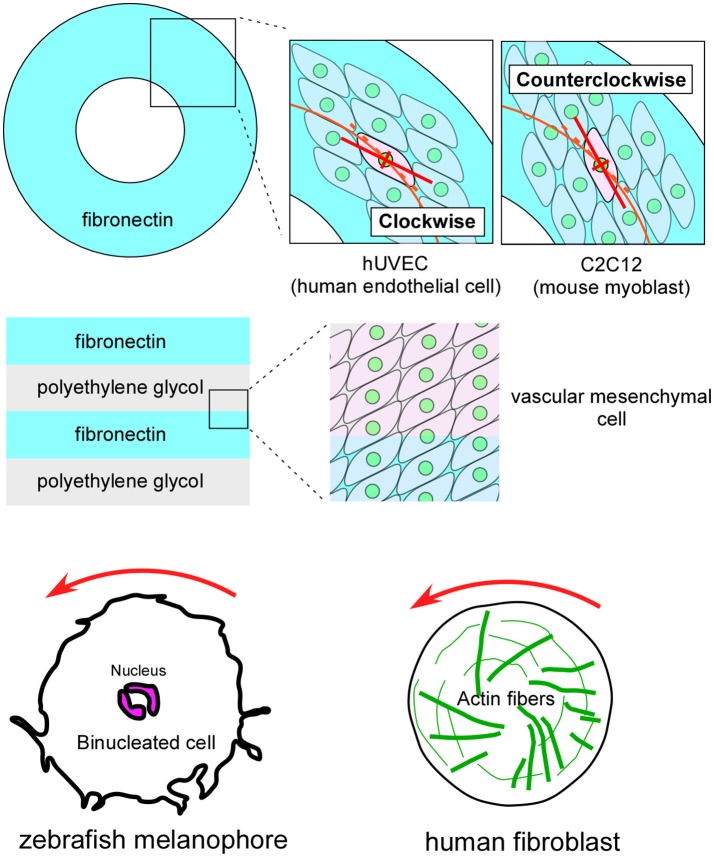
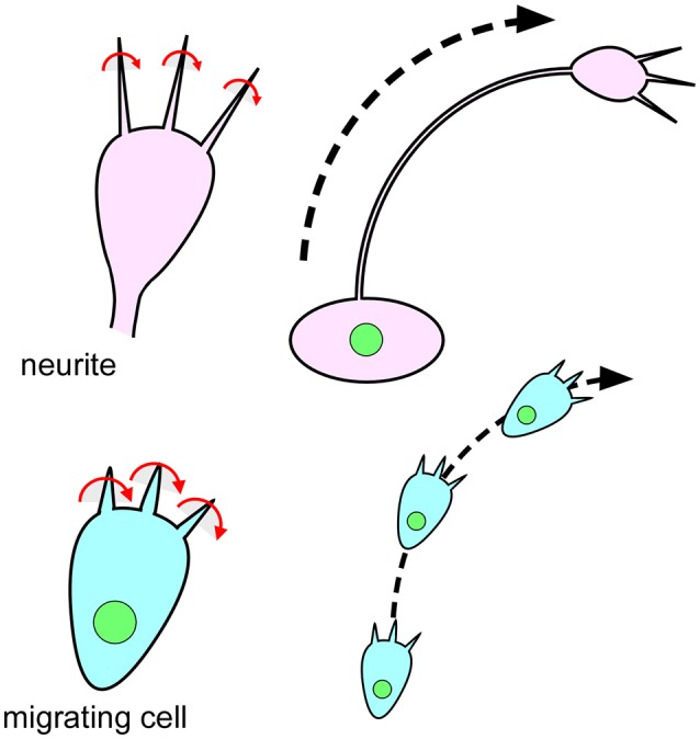
Most macromolecules found in cells are chiral, meaning that they cannot be superimposed onto their mirror image. However, cells themselves can also be chiral, a subject that has received little attention until very recently. In our studies on the mechanisms of left-right (LR) asymmetric development in Drosophila, we discovered that cells can have an intrinsic chirality to their structure, and that this "cell chirality" is generally responsible for the LR asymmetric development of certain organs in this species. The actin cytoskeleton plays important roles in the formation of cell chirality. In addition, Myosin31DF (Myo31DF), which encodes Drosophila Myosin ID, was identified as a molecular switch for cell chirality. In other invertebrate species, including snails and Caenorhabditis elegans, chirality of the blastomeres, another type of cell chirality, determines the LR asymmetry of structures in the body. Thus, chirality at the cellular level may broadly contribute to LR asymmetric development in various invertebrate species. Recently, cell chirality was also reported for various vertebrate cultured cells, and studies suggested that cell chirality is evolutionarily conserved, including the essential role of the actin cytoskeleton. Although the biological roles of cell chirality in vertebrates remain unknown, it may control LR asymmetric development or other morphogenetic events. The investigation of cell chirality has just begun, and this new field should provide valuable new insights in biology and medicine.

Keywords: Drosophila; F-actin; Myosin I; cell chirality; left-right asymmetry.


Figures

Figure 1
Figure 1
The Drosophila embryonic hindgut rotates 90° counterclockwise. (Left) The embryonic hindgut first forms as a bilaterally symmetric structure that curves ventrally (Left). It rotates 90° counterclockwise from the posterior view (Middle), and consequently curves to the right (Right). (Right) The embryonic gut curves rightward at stage 12 in wild-type Drosophila. This figure is partly adapted from Inaki et al. (2016) with permission.
Figure 2
Figure 2
Myosin31DF mutation reverses the cell chirality and hindgut rotation. (Top) A wild-type Drosophila embryo shows normal cell chirality and a rightward-pointing hindgut. (Bottom) In the Myosin31DF mutant, the cell chirality and hindgut laterality are the mirror images of those in its wild-type counterpart. This figure is partly adapted from Inaki et al. (2016) with permission.
Figure 3
Figure 3
A computer simulation recapitulates the cell-chirality-driven counterclockwise rotation of the hindgut. (Top) The shape of the apical surface of hindgut epithelial cells is LR-asymmetric (Left). Considering that these cells also have apical-basal polarity, they show chirality. This property is illustrated by left-handed and right-handed chiral amino acids (Right). (Bottom) To test whether cell chirality alone could induce the axial rotation of the hindgut epithelial tube, a computer simulation based on a vertex model was performed. The introduction of LR bias to the contraction of the cell boundary was sufficient to recapitulate the cell chirality found in vivo. The introduction and subsequent release of cell chirality were sufficient to induce the LR-asymmetric rotation of the model epithelial tube (Taniguchi et al., 2011). These figures are partly adapted from Inaki et al. (2016) and Taniguchi et al. (2011) with permissions.
Figure 4
Figure 4
Cell chirality drives LR asymmetric morphogenesis in various Drosophila organs. (Top) In wild-type Drosophila males, the genitalia rotate clockwise as viewed from the posterior. Just before rotation begins, genital epithelial cells in the A8 segment show a chiral cell shape and an asymmetric Myosin II (MyoII) distribution (Middle). This cell chirality drives the 360° rotation of the genitalia (Right). (Bottom) The wild-type adult Drosophila gut develops from the H2 segment during the pupal stage, while gut laterality is determined by Myo31DF expressed in the H1 segment during the larval stage (Middle). Cell chirality is observed only in the H2 segment after the H1 segment is eliminated during metamorphosis. The handedness determined by Myosin31DF in the H1 segment is propagated to the H2 segment, leading to the LR-directional looping of the adult hindgut (Right). The atypical cadherins Dachsous (Ds) and Fat (Ft) are thought to be involved in this process. Dachsous is reported to bind Myo31DF (Middle). These figures are partly adapted from Inaki et al. (2016) with permission.
Figure 5
Figure 5
Vertebrate cultured cells exhibit intrinsic cell chirality. (Top) Cultured human endothelial (hUVEC) and mouse myoblast (C2C12) cells are arranged in clockwise and counterclockwise spiral patterns on a substrate (Fibronectin) with a ring micropattern (Wan et al., 2011). (Middle) Vascular mesenchymal cells exhibit intrinsic chirality when plated on a substrate with a stripe micropattern (Fibronectin and polyethylene glycol) (Chen et al., 2012). (Bottom) A counterclockwise rotation of the nucleus is observed in cultured zebrafish melanophores (Left) (Yamanaka and Kondo, 2015). Human fibroblasts cultured on a micropattern substrate show chiral swirling (Right) (Tee et al., 2015). These figures are partly adapted from Inaki et al. (2016) with permission.
Figure 6
Figure 6
The chiral behavior of filopodia is evolutionarily conserved from single-celled eukaryotic organisms to mammals. (Top) Filopodia of the growth cone on mouse neurites rotate in a rightward screwlike manner (Left) (Tamada and Igarashi, 2017). The right-screw rotation of filopodia introduces a clockwise growth of neurites (Right) (Tamada and Igarashi, 2017). (Bottom) Filopodia of Dictyostelium cells also show a rightward-screwlike rotation (Left) (Tamada and Igarashi, 2017). The right-screw rotation of filopodia leads the cells to migrate in a clockwise direction (Right) (Tamada and Igarashi, 2017).

References

    1. Bergmann D. C., Lee M., Robertson B., Tsou M. F., Rose L. S., Wood W. B. (2003). Embryonic handedness choice in C. elegans involves the Galpha protein GPA-16. Development 130, 5731–5740. 10.1242/dev.00839 - DOI - PubMed
    1. Brown N. A., Wolpert L. (1990). The development of handedness in left/right asymmetry. Development 109, 1–9. - PubMed
    1. Chen T.-H., Hsu J. J., Zhao X., Guo C., Wong M. N., Huang Y., et al. (2012). Left-right symmetry breaking in tissue morphogenesis via cytoskeletal mechanics novelty and significance. Circ. Res. 110, 551–559. 10.1161/CIRCRESAHA.111.255927 - DOI - PMC - PubMed
    1. Coutelis J. B., Géminard C., Spéder P., Suzanne M., Petzoldt A. G., Noselli S. (2013). Drosophila left/right asymmetry establishment is controlled by the Hox gene abdominal-B. Dev. Cell 24, 89–97. 10.1016/j.devcel.2012.11.013 - DOI - PubMed
    1. Davison A., McDowell G. S., Holden J. M., Johnson H. F., Koutsovoulos G. D., Liu M. M., et al. (2016). Formin is associated with left-right asymmetry in the pond snail and the frog. Curr. Biol. 26, 654–660. 10.1016/j.cub.2015.12.071 - DOI - PMC - PubMed

LinkOut - more resources