Review
. 2018 May;29(5):807-816.
doi: 10.1007/s13361-018-1914-1. Epub 2018 Apr 17.

Diversity of Neuropeptide Cell-Cell Signaling Molecules Generated by Proteolytic Processing Revealed by Neuropeptidomics Mass Spectrometry

Affiliations
Review

Diversity of Neuropeptide Cell-Cell Signaling Molecules Generated by Proteolytic Processing Revealed by Neuropeptidomics Mass Spectrometry

Vivian Hook et al. J Am Soc Mass Spectrom. 2018 May.

Abstract

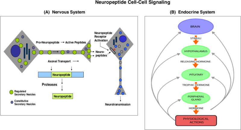
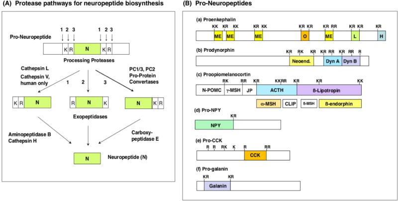
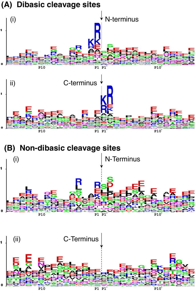
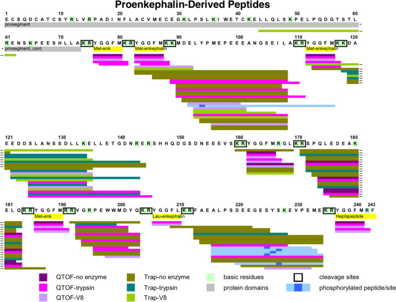
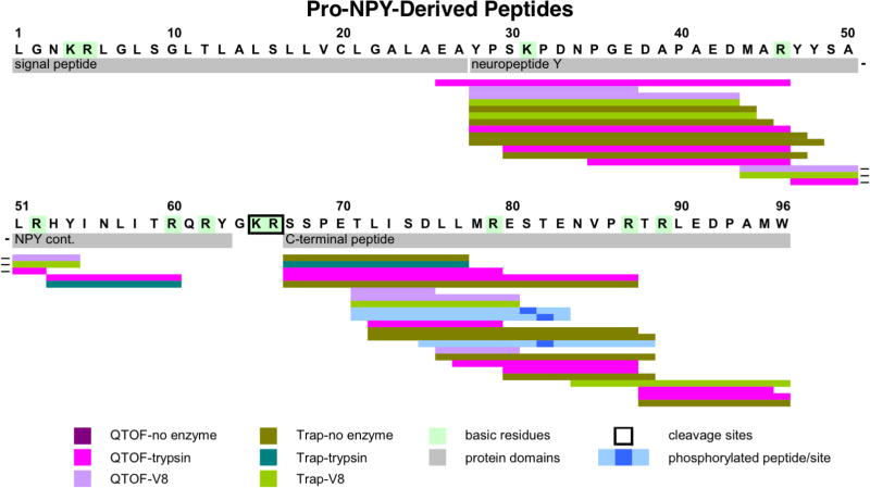
Neuropeptides are short peptides in the range of 3-40 residues that are secreted for cell-cell communication in neuroendocrine systems. In the nervous system, neuropeptides comprise the largest group of neurotransmitters. In the endocrine system, neuropeptides function as peptide hormones to coordinate intercellular signaling among target physiological systems. The diversity of neuropeptide functions is defined by their distinct primary sequences, peptide lengths, proteolytic processing of pro-neuropeptide precursors, and covalent modifications. Global, untargeted neuropeptidomics mass spectrometry is advantageous for defining the structural features of the thousands to tens of thousands of neuropeptides present in biological systems. Defining neuropeptide structures is the basis for defining the proteolytic processing pathways that convert pro-neuropeptides into active peptides. Neuropeptidomics has revealed that processing of pro-neuropeptides occurs at paired basic residues sites, and at non-basic residue sites. Processing results in neuropeptides with known functions and generates novel peptides representing intervening peptide domains flanked by dibasic residue processing sites, identified by neuropeptidomics. While very short peptide products of 2-4 residues are predicted from pro-neuropeptide dibasic processing sites, such peptides have not been readily identified; therefore, it will be logical to utilize metabolomics to identify very short peptides with neuropeptidomics in future studies. Proteolytic processing is accompanied by covalent post-translational modifications (PTMs) of neuropeptides comprising C-terminal amidation, N-terminal pyroglutamate, disulfide bonds, phosphorylation, sulfation, acetylation, glycosylation, and others. Neuropeptidomics can define PTM features of neuropeptides. In summary, neuropeptidomics for untargeted, global analyses of neuropeptides is essential for elucidation of proteases that generate diverse neuropeptides for cell-cell signaling. Graphical Abstract ᅟ.

Keywords: Cathepsin; Endocrine; Enkephalin; Mass spectrometry; NPY; Nervous system; Neuropeptides; Neurotransmission; Peptidomics; Pro-protein convertase; Proteases; Regulation.


Figures

Figure 1
Figure 1. Neuropeptide cell-cell signaling in the nervous and endocrine systems
A. Neuropeptides in the nervous system as peptide neurotransmitters. Neuropeptides are utilized as neurotransmitters for cell-cell communication in the brain and in the peripheral sympathetic and parasympathetic nervous system. Neuropeptides are synthesized within secretory vesicles that are transported from the neuronal cell body to nerve terminals. During axonal transport of such secretory vesicles, pro-neuropeptide precursors (or prohormone) are packaged into newly synthesized secretory vesicles with proteases for precursor processing into mature neuropeptides. Activity-dependent regulation of secretion releases neuropeptides at nerve terminals for neurotransmission. B. Neuropeptides in the endocrine systems as peptide hormones. Neuropeptides function as peptide hormones to mediate cell-cell communication among brain and endocrine systems for regulation of physiological functions. Brain neuropeptides and endocrine peptide hormones are co-regulated in feedback systems for fine tuning of physiological regulation. For example, the hypothalamo-neurophypophyseal system regulates the pituitary-adrenal axis by secretion of CRF from the hypothalamus region of the brain to induce secretion of the ACTH peptide hormone from the pituitary. Released ACTH targets the adrenal cortex for stimulation of glucocorticoid production. Resultant increases in plasma glucocorticoid participate in feedback inhibition of CRF and ACTH to maintain constant levels of glucocorticoid. Numerous peptide hormones regulate physiological functions in stimulatory and feedback system.
Figure 2
Figure 2. Protease Pathways to Convert Pro-Neuropeptides to Neuropeptides
A. Protease pathways for neuropeptide biosynthesis. Pro-neuropeptides undergo proteolytic processing at dibasic residue cleavage sites by dual cysteine and serine subtilisin-like protease pathways. The cathepsin L cysteine protease cleavages pro-neuropeptides at paired basic residues. Resultant peptide intermediates require removal of N-terminal basic residues by aminopeptidase B or cathepsin H, and removal of C-terminal basic residues by carboxypeptidase E (CPE). The human-specific cathepsin V cysteine protease functions only in human cells for neuropeptide production, because the cathepsin V gene is present only in the human genome (and not in the genome of other species of organisms). The serine protease pathway consists of the pro-protein convertases (PC) PC1/3 and PC2 that cleave at paired basic residues. Resultant peptide intermediates then require removal of C-terminal basic residues by CPE. B. Pro-neuropeptide structures. Schematic illustration of the opioid pro-neuropeptide precursors are shown for proenkephalin (PENK), prodynorphin (PDYN), and proopiomelanocortin (POMC), as well as the precursors of neuropeptide Y (NPY), cholecystokinin (CCK), and galanin (GAL). Mature neuropeptides are indicated in colored areas within the pro-neuropeptides. Neuropeptides within the precursors are typically flanked by paired basic residues (Lys-Arg (KR), Lys-Lys(KK), Arg-Lys (RK), and Arg-Arg (RR)), and occasionally at monobasic Arg sites, that are recognized and cleaved by processing proteases to generate mature neuropeptides.
Figure 3
Figure 3. Pro-neuropeptide processing at dibasic and non-dibasic cleavage sites revealed by neuropeptidomics
A. Dibasic residue processing sites. Dibasic residue sites are typical proteolytic cleavage sites of pro-neuropeptides in secretory vesicles. Sequence LOGO maps of the N- and C-termini of neuropeptides in secretory vesicles were generated by evaluation of adjacent residues to the identified peptides with their pro-neuropeptide precursors [7]. (i) N-terminal cleavage sites of identified neuropeptides. The N-termini of peptides are indicated by the arrow, with illustration of the peptide flanking amino acid sequences present within the pro-neuropeptides. The x-axis shows residues at the P1–P15 positions relative to the cleavage sites at P1-↓P1’ residues. The relative frequency of amino acids at each position is illustrated by the sequence LOGO maps. (ii) C-terminal cleavage sites of identified neuropeptides. The C-termini of peptides are indicated by the arrow, with residues of respective pro-neuropeptides flanking the C-termini at P1’–P15’ positions. B. Novel non-dibasic residue processing sites. Non-dibasic residue cleavage sites of pro-neuropeptides are illustrated after removal of the dibasic sites observed in the data of figure 5a. (i) N-terminal cleavage sites. P1–P15 residues flanking the N-termini of identified peptides within their pro-neuropeptides are illustrated. (ii) C-terminal cleavage sites. P1’–P15’ residues flanking the C-termini of identified peptides within their pro-neuropeptide precursors are illustrated. Data for this figure was acquired as reported by Gupta et al., 2010 [7]. Briefly, dense core secretory vesicles from bovine adrenal medulla were isolated in the presence of protease inhibitors, and a low molecular weight peptide pool was prepared by millipore filtration. The sample was subjected to nano-LC-MS/MS peptidomics analyses. The identified peptides were mapped to respective pro-neuropeptide precursors to evaluate proteolytic cleavage sites. These cleavage sites were mapped by LOGO maps.
Figure 4
Figure 4. Neuropeptidomics analyses of proenkephalin-derived peptides without and with trypsin or V8 protease digestion
To identify peptides by neuropeptidomics, the soluble low molecular weight soluble fraction of human chromaffin granules was undigested, or digested with trypsin or V8 protease for nano-LC-MS/MS identification of peptides. Peptides derived from proenkephalin (PE) were mapped to PE, illustrated by colored lines: QTOF-no enzyme (purple), QTOF-trypsin (bright violet), QTOF-V8 (lavender), Trap-no enzyme (olive), Trap-trypsin (turquoise blue), Trap-V8 (bright green). Active enkephalin neuropeptides within PE are shown in yellow, and dibasic cleavage sites are highlighted by boxes. Phosphorylated peptides are indicated (light blue with phosphorylation site indicated by dark blue square). Hyphens at the end of some lines indicate peptides that were split between two lines in the figure.
Figure 5
Figure 5. Neuropeptidomics analyses of proNPY-derived peptides
Peptides derived from human proNPY (NPY, neuropeptide Y) were analyzed by neuropeptidomics of the low molecular weight soluble pool of human chromaffin secretory vesicles without protease digestion, with trypsin, or with V8 protease digestion followed by nano-LC-MS/MS analyses. Peptides derived from proNPY are mapped, illustrated by colored lines: colored lines: QTOF-no enzyme (purple), QTOF-trypsin (bright violet), QTOF-V8 (lavender), Trap-no enzyme (olive), Trap-trypsin (turquoise blue), Trap-V8 (bright green). Phosphorylated peptides are indicated (light blue with phosphorylation site indicated by dark blue square). ProNPY domains of NPY neuropeptide and the C-terminal peptide are indicated. The signal sequence is also shown since one identified peptide apparently included several residues at the C-terminal end of the putative signal sequence.
Figure 6
Figure 6. Regulation of vasoactive neuropeptide profiles by captopril inhibition of angiotensin converting enzyme (ACE)
Plasma neuropeptides in rat were analyzed by neuropeptidomics after administration of captopril, an anti-hypertensive drug inhibitor of the angiotensin converting enzyme (ACE). Neuropeptidomics was assessed in time-course studies by nano-LC-MS/MS with quantitation using stable isotope-labeled internal standards [9]. Separation of peptides by chromatography and multiple reaction monitoring (MRM) quantitated angiotensin I (Ang I), Ang II, Ang1–7, bradykinin 1–8 (BK 1–8), BK-2–9, and kallidin (KD) vasoactive neuropeptides involved in blood pressure regulation. The angiotensin peptides were significantly reduced by the ACE inhibitor, with parallel increases in bradykinins and kallidin (potent vasodilator). The percent change in plasma concentration at each time point after drug administration is shown. These results demonstrate simultaneous profiling of multiple plasma peptides by neuropeptidomics to assess drug-induced changes in vasoactive neuropeptides.

References

    1. Kastin AJ. Handbook of biologically active peptides. Amsterdam: Elsevier; 2006.
    1. Siegel GJ, Albers RW, Fisher SK, Uhler MD. Basic Neurochemistry. Lippincott Williams and Wilkins; Philadelphia: 1999. pp. 363–382.
    1. Hook V, Funkelstein L, Lu D, Bark S, Wegrzyn J, Hwang SR. Proteases for processing proneuropeptides into peptide neurotransmitters and hormones. Annu. Rev. Pharmacol. Toxicol. 2008;48:393–423. - PMC - PubMed
    1. Yin P, Hou X, Romanova EV, Sweedler JV. Neuropeptidomics: mass spectrometry-based qualitative and quantitative analysis. Methods Mol. Biol. 2011;789:223–236. - PMC - PubMed
    1. Ye H, Wang J, Tian Z, Ma F, Dowell JA, Bremer Q, Lu G, Baldo B, Li L. Quantitative mass spectrometry reveals food intake-induced neuropeptide level changes in rat brain: functional assessment of selected neuropeptides as feeding regulators. Mol. Cell Proteomics. pii: mcp.000057.2017. - PMC - PubMed