

Cone synapses in mammalian retinal rod bipolar cells
- PMID: 29667170
- PMCID: PMC6031453
- DOI: 10.1002/cne.24456
Cone synapses in mammalian retinal rod bipolar cells
Abstract
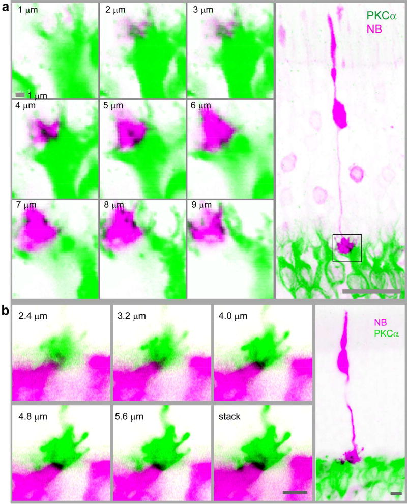
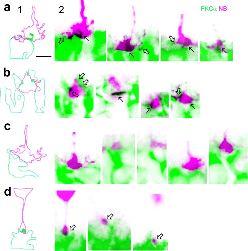
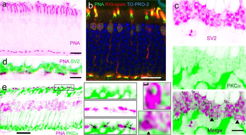
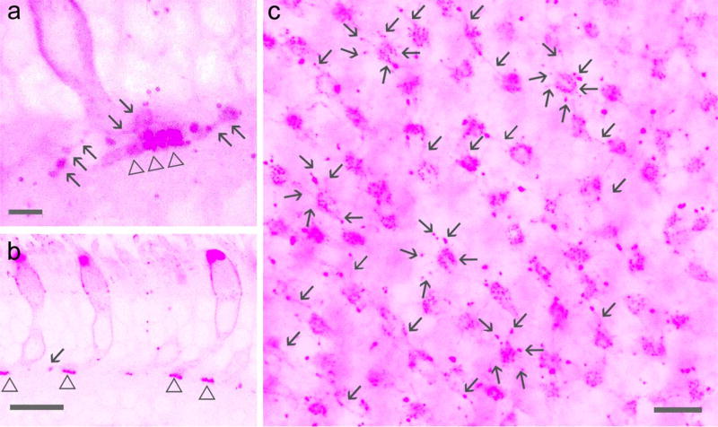
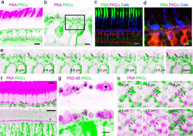
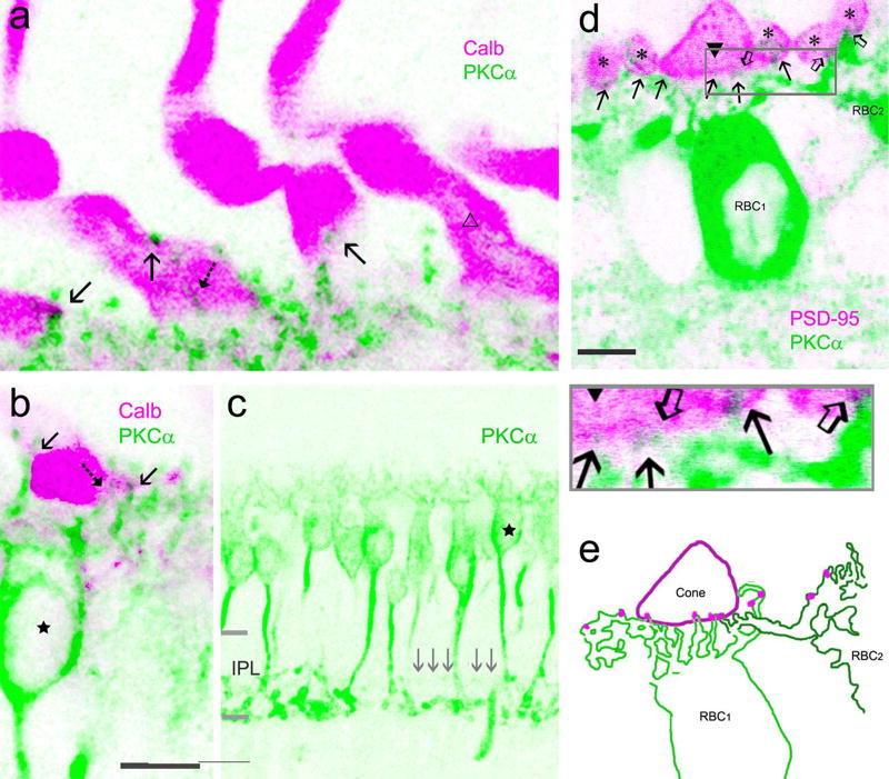
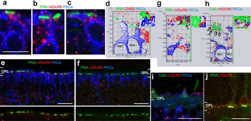
Some mammalian rod bipolar cells (RBCs) can receive excitatory chemical synaptic inputs from both rods and cones (DBCR2 ), but anatomical evidence for mammalian cone-RBC contacts has been sparse. We examined anatomical cone-RBC contacts using neurobiotin (NB) to visualize individual mouse cones and standard immuno-markers to identify RBCs, cone pedicles and synapses in mouse and baboon retinas. Peanut agglutinin (PNA) stained the basal membrane of all cone pedicles, and mouse cones were positive for red/green (R/G)-opsin, whereas baboon cones were positive for calbindin D-28k. All synapses in the outer plexiform layer were labeled for synaptic vesicle protein 2 (SV2) and PSD (postsynaptic density)-95, and those that coincided with PNA resided closest to bipolar cell somas. Cone-RBC synaptic contacts were identified by: (a) RBC dendrites deeply invaginating into the center of cone pedicles (invaginating synapses), (b) RBC dendritic spines intruding into the surface of cone pedicles (superficial synapses), and (c) PKCα immunoreactivity coinciding with synaptic marker SV2, PSD-95, mGluR6, G protein beta 5 or PNA at cone pedicles. One RBC could form 0-1 invaginating and 1-3 superficial contacts with cones. 20.7% and 38.9% of mouse RBCs contacted cones in the peripheral and central retina (p < .05, n = 14 samples), respectively, while 34.4% (peripheral) and 48.5% (central) of cones contacted RBCs (p > .05). In baboon retinas (n = 4 samples), cone-RBC contacts involved 12.2% of RBCs (n = 416 cells) and 22.5% of cones (n = 225 cells). This suggests that rod and cone signals in the ON pathway are integrated in some RBCs before reaching AII amacrine cells.
Keywords: PKCα RRID: AB_477345; Postsynaptic density-95 RRID: AB_10807979; RRID: AB_2313718; RRID: AB_2650488; cone synapse; confocal microscopy; immunohistology; mGluR6 RRID: AB_784501; mouse; primate; rod bipolar cell; synaptic vesicle protein RRID:AB_2315387; tracer injection.
© 2018 Wiley Periodicals, Inc.
Conflict of interest statement
Conflict of interest statement
All authors do not have any known or potential conflict of interest including financial, personal or other relationships with other people or organizations within three years of beginning the submitted work that could inappropriately influence, or be perceived to influence, their work.
Figures
References
-
- Aartsen WM, Kantardzhieva A, Klooster J, van Rossum AG, van de Pavert SA, Versteeg I, et al. Mpp4 recruits Psd95 and Veli3 towards the photoreceptor synapse. Hum. Mol. Genet. 2006;15:1291–1302. - PubMed
-
- Applebury ML, Antoch MP, Baxter LC, Chun LL, Falk JD, Farhangfar F, et al. The murine cone photoreceptor: a single cone type expresses both S and M opsins with retinal spatial patterning. Neuron. 2000;27:513–523. - PubMed
-
- Bajjalieh SM, Peterson K, Shinghal R, Scheller RH. SV2, a brain synaptic vesicle protein homologous to bacterial transporters. Science. 1992;257:1271–1273. - PubMed
Publication types
MeSH terms
Grants and funding
LinkOut - more resources
Full Text Sources
Other Literature Sources
Miscellaneous
