Review
. 2018 May;22(5):419-437.
doi: 10.1080/14728222.2018.1465931. Epub 2018 May 7.

Emerging therapeutic targets for neurofibromatosis type 1

Affiliations
Review

Emerging therapeutic targets for neurofibromatosis type 1

James A Walker et al. Expert Opin Ther Targets. 2018 May.

Abstract

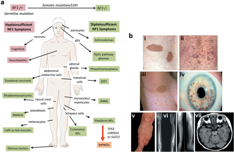
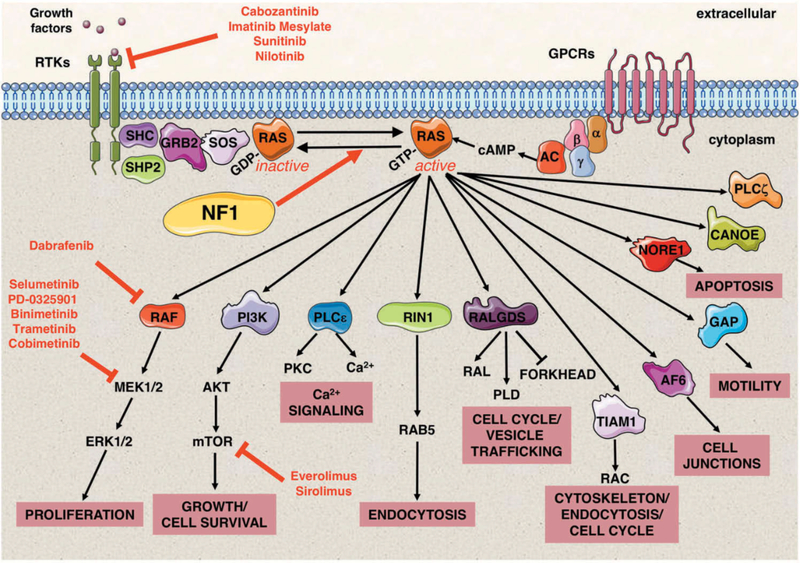
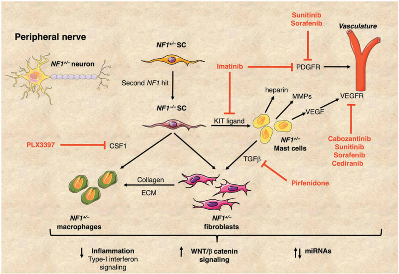
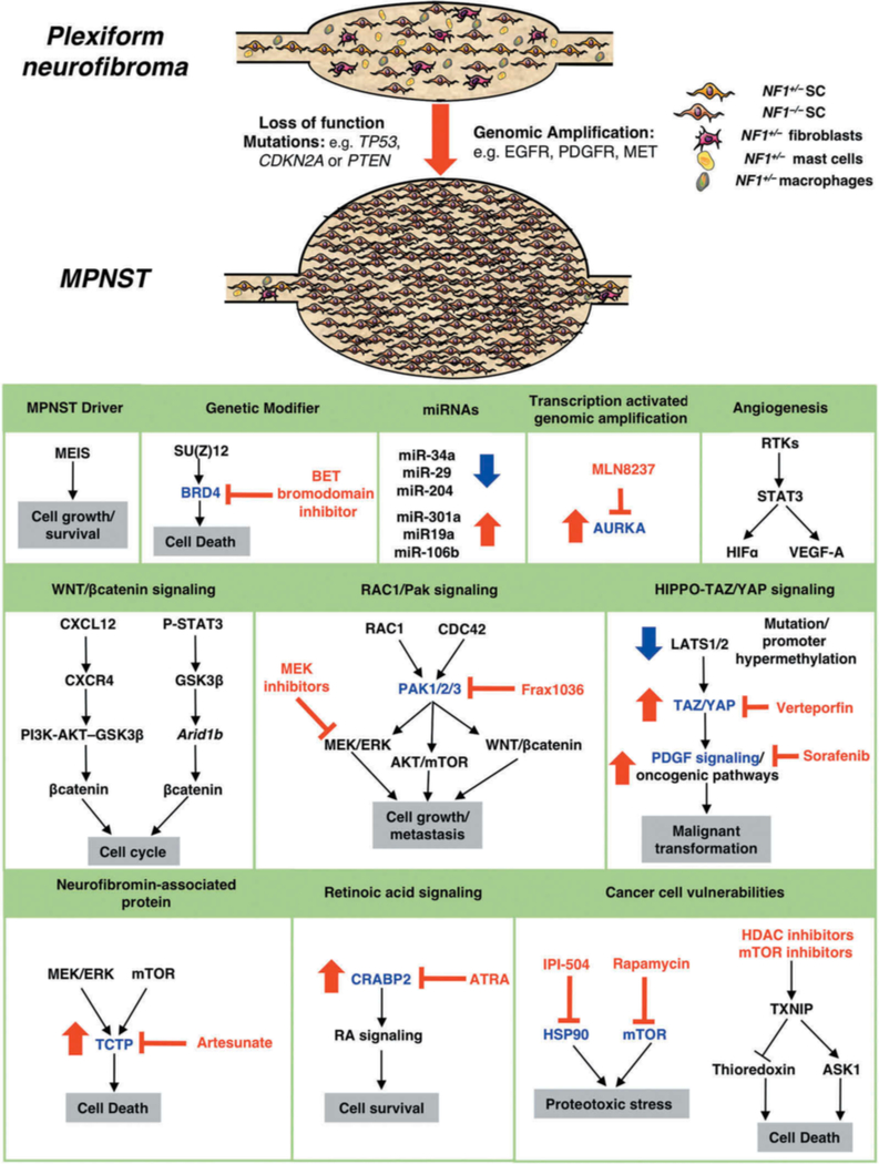
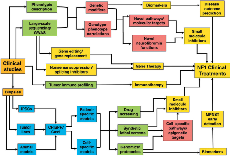
Neurofibromatosis type 1 (NF1) is an autosomal dominantly inherited tumor predisposition syndrome with an incidence of one in 3000-4000 individuals with no currently effective therapies. The NF1 gene encodes neurofibromin, which functions as a negative regulator of RAS. NF1 is a chronic multisystem disorder affecting many different tissues. Due to cell-specific complexities of RAS signaling, therapeutic approaches for NF1 will likely have to focus on a particular tissue and manifestation of the disease. Areas covered: We discuss the multisystem nature of NF1 and the signaling pathways affected due to neurofibromin deficiency. We explore the cell-/tissue-specific molecular and cellular consequences of aberrant RAS signaling in NF1 and speculate on their potential as therapeutic targets for the disease. We discuss recent genomic, transcriptomic, and proteomic studies combined with molecular, cellular, and biochemical analyses which have identified several targets for specific NF1 manifestations. We also consider the possibility of patient-specific gene therapy approaches for NF1. Expert opinion: The emergence of NF1 genotype-phenotype correlations, characterization of cell-specific signaling pathways affected in NF1, identification of novel biomarkers, and the development of sophisticated animal models accurately reflecting human pathology will continue to provide opportunities to develop therapeutic approaches to combat this multisystem disorder.

Keywords: Neurofibromas; RAS-MEK-ERK signaling; RASopathies; neurofibromatosis type 1; peripheral nerve sheath tumors.


Figures

Figure 1
Figure 1. Neurofibromatosis type 1 (NF1) is a multisystem disorder.
A. NF1 patients are predisposed to developing symptoms affecting multiple cells of origin and tissues. Some manifestations associated with NF1, such as cognitive and vascular problems, result from haploinsufficiency of NF1. In contrast, other symptoms are triggered by somatic NF1 mutation/loss of heterozygosity (LOH) resulting in biallelic NF1 inactivation. Further, transformation of plexiform neurofibromas (NFs) into malignant peripheral nerve sheath tumors (MPNSTs) involves additional genetic events. Abbreviations: juvenile myelomonocytic leukemias (JMML) and gastrointestinal stromal tumors (GIST). B. The major defining features of NF1 include: (i) Café-au-lait macules, (ii) cutaneous neurofibromas, (iii) axillary freckling, (iv) Lisch nodules, (v) plexiform neurofibromas, (vi) thinning of long bone cortex and (vii) optic pathway glioma. Adapted from [2].
Figure 2
Figure 2. Neurofibromin is a negative regulator of the RAS signaling pathway
RAS proteins function as fundamental signaling switches controlling a multitude of cellular processes including normal cell growth and differentiation. RAS proteins exist in two cellular states: an inactive GDP-bound form and an active GTP-bound form. Although RAS has a low intrinsic GTPase-activity, RAS-GTPase activating proteins (GAPs) stimulate this activity and consequently help to hydrolyze bound GTP back to GDP. Conversely, guanine nucleotide exchange factors (GEFs), e.g. SOS, convert RAS to its active GTP-bound form. NF1 encodes neurofibromin, a RAS-GAP, which is able to negatively regulate HRAS, KRAS, NRAS, MRAS, RRAS, and RRAS2 (TC21). Neurofibromin deficiency therefore results in elevated RAS signaling. RAS signaling transduces extracellular signals from ligand-activated receptors, both receptor tyrosine kinases (RTKs) and G protein-coupled receptors (GPCRs) at the cell surface through multiple pathways. RAS-GTP proteins activate a multitude of effector proteins, including RAF proteins (ARAF, BRAF, and CRAF) and the MEK–ERK signaling cascade, RalGDS, and the PI3 kinase family. The molecular and cellular events controlled by normal and aberrantly regulated RAS signaling are cell-type specific, depending on the expression of different RAS isoforms and their relative engagement of particular RAS effector proteins. Small molecule inhibitors to upstream regulators of RAS and its downstream effectors in NF1 clinical trials are shown in red. Abbreviations: Growth factor receptor-bound protein 2 (GRB2), src homology 2 domain (SHC), adenyl cyclase (AC), cyclic adenosine monophosphate (cAMP), α, β and γ represent the subunits of a heterotrimeric G protein complex.
Figure 3
Figure 3. Plexiform neurofibromas in NF1
Plexiform neurofibromas (PNFs) develop from NF1+/− Schwann cells (SC) with a second hit resulting in NF1 biallelic inactivation (NF1−/−). These tumors comprise a mixture of SCs, mast cells, macrophages and fibroblasts intrinsic to the peripheral nerve. The growth of PNFs depends on the complex interplay between these cell types. KIT ligand is secreted by NF1−/− SCs and acts as a chemo-attractant for NF1+/− mast cells. In turn, NF1+/− mast cells produce TGFβ, stimulating NF1+/− fibroblasts to increase collagen production and other extracellular matrix (ECM) proteins. NF1+/− mast cells also produce heparin, vascular endothelial growth factor (VEGF) and matrix metalloproteinases (MMPs) promoting tumor vascularization and tumor invasiveness. NF1−/− SCs secrete colony-stimulating factor (CSF1) thereby recruiting macrophages, aiding tumor progression. Small molecule inhibitors in NF1 clinical trials are shown in red. Recent reports have highlighted the importance of inflammation, increased signaling through the WNT/β-catenin pathway and misregulated miRNAs in PNFs, all of which may represent potential therapeutic targets.
Figure 4
Figure 4. Progression of plexiform neurofibromas to malignant peripheral nerve sheath tumors
MPNSTs are highly invasive soft tissue sarcomas that frequently metastasize and arise from benign PNFs harboring biallelic NF1 inactivation. Additional genetic changes leading to the transition from PNFs to MPNSTs include loss-of-function mutations in tumor suppressors such as TP53, CDKN2A and PTEN, or genomic amplification of RTKs or signaling factors. NF1−/− SCs consequently have dramatically increased proliferation rates compared to those in benign PNFs. Potential therapeutic targets for MPNSTs are highlighted. Target proteins implicated in the malignant progression of MPNSTs are shown in blue (p21-activated kinases (PAK1/2/3), bromodomain-containing protein 4 (BRD4), Cellular Retinoic Acid Binding Protein 2 (CRABP2), heat shock protein 90 (HSP90), mTOR, Translationally controlled tumor protein (TCTP) and Aurora kinase A (AURKA)) and small molecule inhibitors (BET bromodomain inhibitors, FRAX1036, IPI-504, rapamycin, HDAC inhibitors, artesunate, MLN8237, all-trans retinoic acid (ATRA)) in red. Increase in level of protein/miRNA in MPNSTs is denoted by red arrow, blue arrow denotes decrease.
Figure 5
Figure 5. Network of approaches leading to discovery and testing of new therapeutic targets in NF1
1) Clinical examination of patients combined with molecular analyses is beginning to reveal NF1 genotype-phenotype correlations - findings that may define novel functions of neurofibromin and identify new therapeutic targets. In addition, genome-wide association studies (GWAS) offer a platform to identify genetic modifiers, which will facilitate the identification of novel targets and biomarkers. Understanding the effect of environmental factors and hormones on NF1 disease progression may also reveal novel treatments. 2) Patient-derived biopsies will aid in the generation of: a) induced pluripotent stem cells (iPSCs) to allow development of patient and cell-specific models and give insights for new targets and biomarkers for different NF1 clinical manifestations; b) tumor-specific cell lines and animal models accurately reflecting the human disease that will permit improved screening of small molecule inhibitors; c) patient-derived cell line models will facilitate cellular pathway analysis (in particular RAS pathway and its upstream and downstream effectors (including receptor tyrosine kinases and micro RNAs) identification of therapeutic targets and biomarkers, and will also allow the testing of novel drugs including small molecule inhibitors prior to their use in clinical trials; d) synthetic lethal screening (using CRISPR libraries) could be exploited to devise therapies to selectively kill NF1-deficient tumors; e) immune profiling leading to immunotherapy and generation of novel biomarkers for NF1-associated tumors. 3) Gene therapy approaches focus on antisense oligonucleotides (ASOs) and nonsense suppression, whereas potential correction of mutations via gene editing offers a possibility of restoring endogenous NF1 gene function, thereby providing a long-term solution for NF1 patients. Highlighted boxes: clinical studies/biopsies (orange), genetic analysis and screening (green), disease models (blue), new insights into basic NF1 biology (red), potential therapeutic approaches and clinical treatments (yellow).

References

    1. Upadhyaya M, Cooper DN. “Molecular and Cellular Biology of Neurofibromatosis Type 1”. 2012:Eds: Upadhyaya M and Cooper DN Springer Verlag.
    1. Upadhyaya M Neurofibromatosis type 1: diagnosis and recent advances. Expert Opin Med Diagn. 2010. July;4(4):307–22. doi: 10.1517/17530059.2010.494660. - DOI - PubMed
    1. Evans DG, Baser ME, McGaughran J, et al. Malignant peripheral nerve sheath tumours in neurofibromatosis 1. J Med Genet. 2002. May;39(5):311–4. - PMC - PubMed
    1. Farid M, Demicco EG, Garcia R, et al. Malignant peripheral nerve sheath tumors. Oncologist. 2014. February;19(2):193–201. doi: 10.1634/theoncologist.2013-0328. - DOI - PMC - PubMed
    1. Gutmann DH, Ferner RE, Listernick RH, et al. Neurofibromatosis type 1. Nat Rev Dis Primers. 2017. February 23;3:17004. doi: 10.1038/nrdp.2017.4. - DOI - PubMed

Publication types